

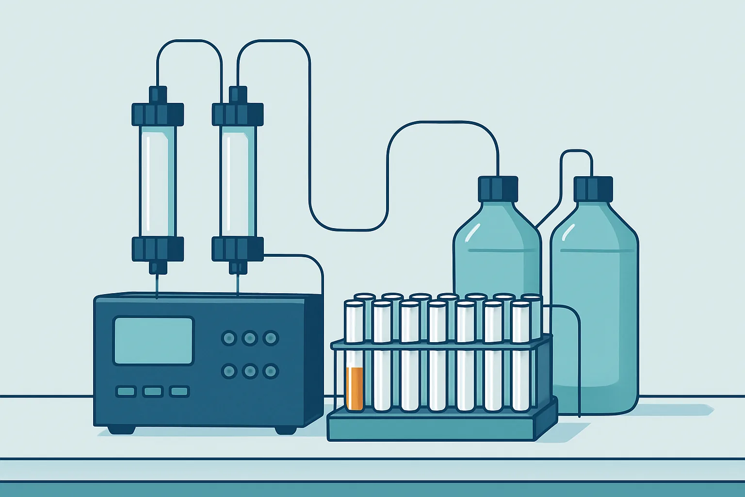
Most purification failures are not technique failures. The columns run, the buffers are correct, the gel is stained properly. The protein simply is not there in the amount or the state that the next experiment needs, and by the time that becomes obvious the material has been through four steps and cannot be recovered.
The reason is almost always that the strategy was assembled rather than designed. Someone picks a tag because it is what the lab has, adds a gel filtration step at the end because that is what people do, and discovers late that the protein aggregates, or co-purifies with a chaperone, or has lost activity somewhere between lysis and storage.
A purification is a sequence of separations, each exploiting a different physical property. The useful mental model is orthogonality: every step should sort molecules on a basis that the previous step ignored. Get that right and three steps can take you from a cell paste to something usable. Get it wrong and six steps will still leave contaminants that travelled with your protein the whole way.
Key takeaways
- Define what purity means for your specific downstream application before choosing any column.
- Steps should be orthogonal, separating on charge, size, hydrophobicity or specific binding in turn.
- Yield is lost at every step, so fewer well-chosen steps usually beat more mediocre ones.
- Affinity tags are convenient but change the protein, and their position and removal need deciding early.
- Track protein amount and activity at each step, not just at the end, or you cannot tell where things went wrong.
Defining Purity for Your Application
Purity is not a single number, and treating it as one is the most common planning error. A protein that is entirely adequate for raising antibodies may be useless for crystallography, and a preparation that crystallises beautifully may be unacceptable for a cell-based assay because of residual endotoxin.
Work backwards from what the protein must do. For structural work the requirement is chemical and conformational homogeneity: a single species, one oligomeric state, no partial proteolysis, minimal buffer heterogeneity. For enzyme kinetics the requirement is that no contaminating activity competes with or acts on your substrate, which can matter even at trace levels invisible on a gel. For antibody production the requirement is that no contaminant is immunogenic enough to dominate the response. For therapeutic or cell-culture use, host cell proteins, nucleic acid and endotoxin all carry limits independent of what a stained gel shows.
This matters because it determines what you measure. A coomassie-stained gel showing one band is a low bar; it typically cannot resolve contaminants present below roughly one percent of the total. Silver staining goes further. Analytical size exclusion tells you about aggregation and oligomeric state, which a denaturing gel destroys and therefore never reveals. Mass spectrometry tells you about proteolysis and modification. Each answers a different question, and picking the wrong readout means declaring success on the wrong criterion.
The other half of the definition is quantity. Purity and yield trade against each other continuously, and a preparation that is exquisitely pure but yields too little to run the experiment has failed just as completely as a dirty one.
Cell Lysis and Initial Clarification
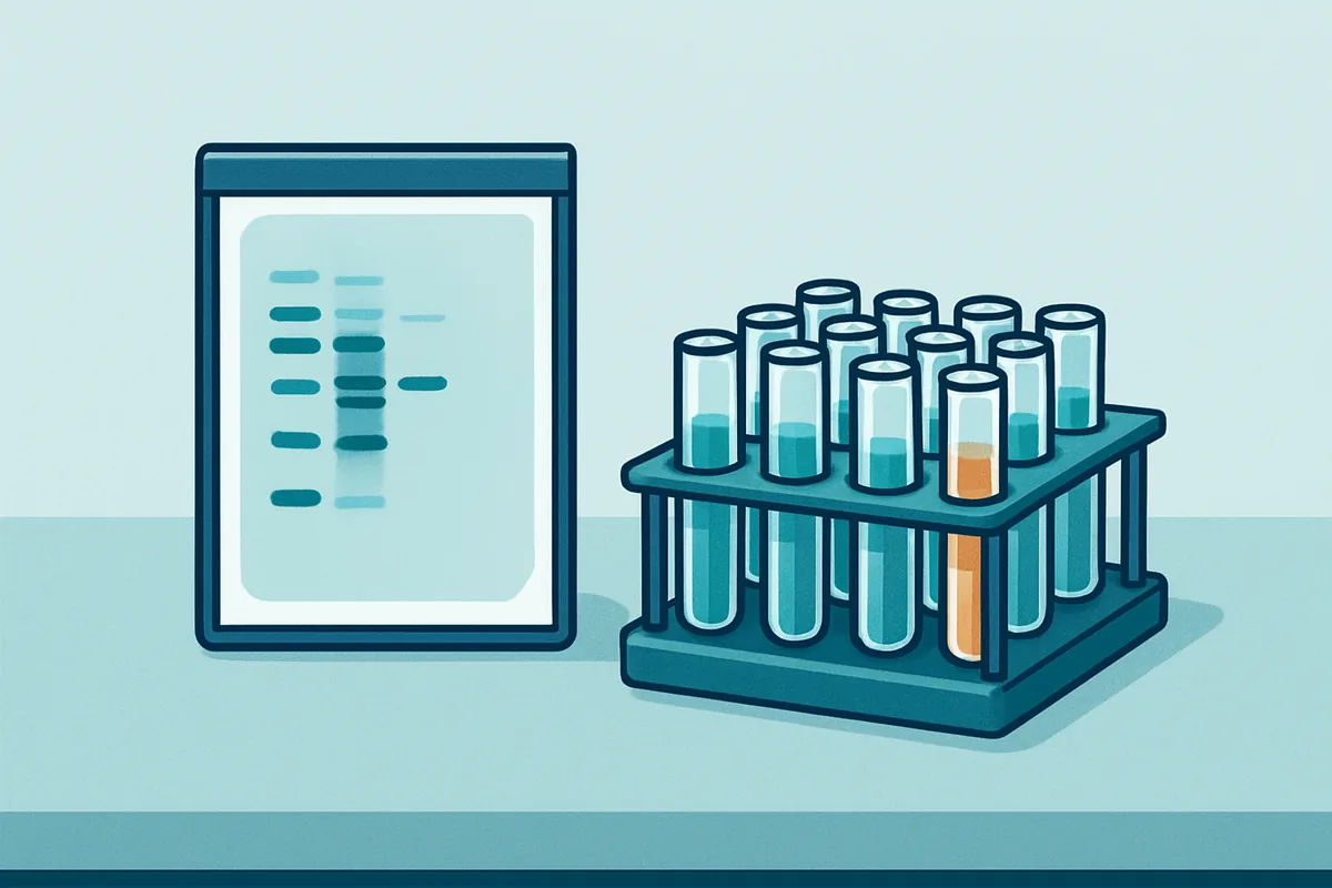
Everything downstream inherits the condition of the lysate, and lysis is where most irreversible damage happens.
The mechanical problem is straightforward: break the cell without breaking the protein. Sonication is fast and cheap but deposits heat locally and generates shear and free radicals, all of which denature sensitive proteins; it works well for small volumes with careful cooling and pulsing. High-pressure homogenisation is more consistent and scales better. Enzymatic lysis with lysozyme is gentle but incomplete on its own and usually needs a freeze-thaw or detergent to finish the job. Bead beating suits organisms with tough walls, including yeast and many bacteria, at the cost of considerable heat.
The chemical problem is that lysis releases everything at once, including proteases that were previously compartmentalised away from your protein. From the moment the cell breaks, proteolysis is running. Keeping the lysate cold slows it, protease inhibitors slow it further, and speed does the rest. A lysate that sits on the bench for an hour while someone hunts for a column is a lysate that has already lost its intact termini.
Nucleic acid is the other immediate issue. Released genomic DNA makes the lysate viscous, which fouls filters and columns and traps protein. A nuclease treatment or additional shearing resolves it, and doing so before clarification saves considerable trouble later.
Clarification itself is centrifugation, filtration, or both. High-speed centrifugation removes membranes and debris; a subsequent filtration step protects the first column from the fines that centrifugation leaves behind. Skipping filtration is a false economy, since a blocked column costs more than a filter.
Affinity Tags and Their Trade-Offs
Affinity capture is the highest-resolution step available, because it separates on a specific interaction rather than a bulk property. A single well-behaved affinity step can take a crude lysate to substantially pure protein in one pass, which is why it usually goes first.
The polyhistidine tag is the default for good reasons: it is small, it works under denaturing conditions, the resin is inexpensive, and elution with imidazole or a pH shift is simple. Its weakness is specificity. Host proteins with surface histidine clusters bind the resin too, and a histidine tag on an abundantly expressed protein will still deliver a visible background. Raising the imidazole concentration in the wash reduces that background at some cost in yield, and the correct level has to be found empirically.
Larger tags behave differently. Glutathione S-transferase and maltose-binding protein both frequently improve solubility, which can rescue a protein that otherwise ends up in inclusion bodies, but they are substantial proteins in their own right and often must be removed before any structural or functional work. Streptavidin-based tags offer very high specificity and gentle elution, at higher resin cost.
| Approach | Typical strength | Main limitation | Elution |
|---|---|---|---|
| Polyhistidine | Small, cheap, works when denatured | Host protein background | Imidazole or low pH |
| Glutathione S-transferase | Often aids solubility | Large; dimerises | Reduced glutathione |
| Maltose-binding protein | Strong solubility benefit | Large; must usually be cleaved | Maltose |
| Streptavidin-family | High specificity, gentle | Higher resin cost | Competing ligand |
| Tag-free, native properties | No modification at all | Must be developed per protein | Depends on method |
Two decisions need making before expression, not after. The first is tag position: an N-terminal tag can interfere with signal sequences or with folding of the N-terminal domain, while a C-terminal tag reports on full-length translation because truncated products lose it. The second is removal. If the tag must go, a protease site has to be designed in, and cleavage adds a step with its own yield loss and requires separating the freed tag, the protease and any uncleaved material. Deciding this after the construct exists usually means making the construct again.
Ion Exchange and Charge-Based Separation
Ion exchange separates on net surface charge, which is a property essentially unrelated to specific binding. That makes it a natural partner to affinity capture.
The principle is that a protein’s net charge depends on the pH of the buffer relative to its isoelectric point. Above the isoelectric point the protein carries net negative charge and binds a positively charged anion exchange resin; below it, the reverse. Choosing the buffer pH is therefore choosing which proteins bind at all, and a shift of one pH unit can completely change the separation.
In practice the most useful property of ion exchange is not the binding but the elution. Bound protein is released by a gradually rising salt concentration, and proteins release in order of how strongly they were held. This gives genuine resolution: contaminants that bound alongside your protein often elute at measurably different salt concentrations, and a shallow gradient across the relevant range separates them. A step elution is faster and gives less resolution, which is a reasonable trade when the contaminants are far away in binding strength.
Ion exchange also concentrates. Protein loaded from a large dilute volume elutes in a small one, which is often worth more than the purification itself when the preceding step produced litres of dilute material.
The practical constraint is that the sample must be in low salt to bind, which frequently means diluting or exchanging buffer after an affinity elution. That intermediate handling step is where dilute proteins tend to precipitate, and it is worth planning rather than improvising.
Hydrophobic interaction chromatography is the mirror image and pairs well with it: it binds in high salt and elutes in low, so a high-salt ion exchange eluate can be loaded directly.
Size Exclusion as a Polishing Step
Size exclusion separates by hydrodynamic size, with larger molecules excluded from the pores of the beads and eluting first. It is the least efficient step in the sequence by loading capacity, because resolution depends on the sample occupying only a small fraction of the column volume, and it dilutes rather than concentrates. It goes last for both reasons.
What justifies it is that it answers questions no other step does. It separates aggregates from correctly folded monomer, which matters enormously for anything downstream that is sensitive to oligomeric state. It resolves an oligomeric assembly from its dissociated components. It removes small molecules completely, so it doubles as a buffer exchange into the final storage conditions. And the shape of the elution profile is itself data: a broad, tailing or shouldered peak is telling you about heterogeneity that a gel will not show.
Resolution here is limited. Two proteins need a substantial size difference before a preparative column will separate them cleanly, so size exclusion is a poor choice for removing a contaminant of similar mass. Loading a small volume of concentrated sample onto a long column is what makes it work, which is the opposite of how the earlier steps are loaded.
Tracking Yield and Purity Across Steps
The single habit that most improves purification is keeping a table. Measure total protein and total activity, if there is an assay, at every stage: lysate, clarified lysate, flow-through, wash, eluate, and each subsequent step.
From those numbers come two derived quantities. Specific activity, meaning activity per unit of total protein, should rise at every purification step; if it does not, that step removed nothing useful. Recovery, meaning the fraction of the previous step’s activity that survived, shows where material is being lost. Together they distinguish the two failure modes, because a step can fail by removing nothing or by destroying your protein, and the remedies are opposite.
Saving samples matters as much as measuring them. Keep a small aliquot of every fraction, including the ones you expect to discard, and run them together on one gel at the end. The flow-through that “should” contain nothing is where an unbound protein reveals itself, and once discarded that information is gone.
The most common discovery from this discipline is that a step everyone assumed was essential contributes nothing measurable, or that most of the loss happens in a single handling operation such as a concentration or dialysis. Both findings change the strategy far more than optimising a gradient would.
Storage, Buffer Exchange and Stability
A purified protein is a metastable object, and the buffer it sits in determines how long it stays that way.
Buffer composition does several jobs. The buffering species holds pH near the protein’s stability optimum, which is not necessarily physiological. Salt screens surface charges and prevents both aggregation and non-specific binding to surfaces. Where the protein has free cysteines, a reducing agent prevents intermolecular disulphide formation, but reducing agents oxidise over time and need replacing. Glycerol is widely used because it stabilises the folded state and suppresses ice crystal formation on freezing.
Concentration cuts both ways. Very dilute protein adsorbs to container walls and loses activity, and can be lost entirely at low microgram amounts. Very concentrated protein aggregates. The workable range differs by protein and is worth establishing rather than assuming.
Freezing is where a great deal of carefully purified material dies. Ice formation concentrates solutes in the remaining liquid, exposing the protein to salt and pH conditions far from those in the tube, and the freeze-thaw interface itself denatures many proteins. Freezing rapidly in small single-use aliquots addresses both problems, because it shortens the time spent in the damaging intermediate state and removes any reason to thaw the same tube twice. A cryoprotectant helps further.
Whatever the storage decision, test it. Take one aliquot, freeze it, thaw it and measure activity against fresh material. That comparison takes an afternoon and tells you whether the past two weeks of work will still be usable next month, which is a question worth answering before you need the answer.
Frequently asked questions
Should affinity chromatography always come first?
Usually, because it gives the largest purification factor in a single step and reduces the volume dramatically, which makes everything afterwards easier. The exceptions are when the crude lysate would foul or overload the resin, or when the target is a small fraction of a very large volume, in which case a cheap bulk capture step such as ion exchange or a precipitation first can protect an expensive affinity resin.
How do I decide whether to remove the tag?
Ask whether the tag can plausibly interfere with the intended use. For structural work, functional assays involving the tagged terminus, or any application where the tag’s own properties might dominate, remove it. For pull-downs, immobilisation or antibody production, it can often stay. Removal costs a protease step, a subtractive separation, and some yield, so it should be a decision rather than a default in either direction.
Why does my protein look pure on a gel but behave badly downstream?
A denaturing gel reports the mass of polypeptides present. It says nothing about folding, oligomeric state, aggregation or bound ligands, and it cannot see contaminants below roughly one percent. A single band is entirely compatible with a sample that is half soluble aggregate, or that has lost a cofactor, or that carries a trace contaminating enzyme. Analytical size exclusion and an activity measurement answer questions the gel cannot.
Is more purification steps always better?
No. Every step loses material, and the losses compound, so a four-step protocol recovering three-quarters at each stage delivers under a third of what you started with. Adding a step is only worth it if it removes something that actually interferes with the downstream application. This is why defining purity requirements first matters so much: it tells you when to stop.
What should I do if the protein ends up in inclusion bodies?
There are three broad options. Change the expression conditions, since lower temperature, slower induction, a different host strain or co-expressed chaperones frequently shift the balance toward soluble protein. Add a solubility-enhancing fusion partner. Or purify the inclusion bodies, which are often quite pure to begin with, solubilise them under denaturing conditions and refold. Refolding is protein-specific and empirical, so the first two options are usually tried first.
The strategy that works is drafted before any column is packed. Write down what the protein must do at the end, choose the smallest number of orthogonal steps that plausibly gets there, decide the tag and its fate at construct design, and set up the measurement table before the first lysis. Purification rewards planning far more than it rewards technique, and the labs that reliably get usable protein are not the ones with better hands. They are the ones that decided what they were doing before they started.



