

Two things about coagulation testing surprise almost everyone who looks into it properly. The first is that the tests do not measure clotting as it happens in the body; they measure how fast plasma clots in a tube after being deliberately pushed down one particular pathway with a reagent. The second is that the INR, the number that governs warfarin dosing worldwide, is not a measurement at all. It is a mathematical correction invented so that a result produced in one laboratory would mean the same thing as a result produced in another.
That second point explains a great deal of otherwise confusing behaviour. It explains why the INR is quoted for warfarin and not for other situations, why a prothrombin time in seconds is nearly uninterpretable across laboratories, and why an INR measured on a patient not taking a vitamin K antagonist should be treated with caution.
Key takeaways
- Both PT and aPTT measure the time to clot formation in plasma after a specific pathway is deliberately triggered.
- The INR is a correction that maps a laboratory’s PT onto a common scale using a reagent-specific sensitivity index.
- The INR is validated for stable warfarin therapy and is unreliable in liver disease, acute illness and other anticoagulation.
- Citrate tubes must be filled to the line, because the anticoagulant-to-plasma ratio is part of the test.
- Direct oral anticoagulants affect these tests inconsistently, so a normal PT or aPTT does not exclude their presence.
The Clotting Cascade a Lab Actually Tests
The textbook cascade, with an intrinsic limb, an extrinsic limb and a common final path, is a laboratory construct rather than a description of physiology. It survives because it maps neatly onto the two screening tests, and it should be understood as a model of those tests rather than of a bleeding wound.
In the body, clotting begins when tissue factor, normally sequestered away from flowing blood, becomes exposed by injury. It binds factor VII, and that complex activates factor X, which converts prothrombin to thrombin. The small initial burst of thrombin then amplifies the whole process by activating other factors and platelets, and the contact factors of the so-called intrinsic pathway contribute to sustaining that amplification rather than starting anything. The initiating event is the same regardless of which screening test is being performed.
The laboratory divides this artificially. The prothrombin time bypasses the beginning by adding tissue factor directly, so it interrogates factor VII and then the common path of factors X and V, prothrombin and fibrinogen. The activated partial thromboplastin time triggers the contact system instead, so it interrogates factors XII, XI, IX and VIII before joining the same common path.
Two consequences follow, and they are the ones worth remembering. Because both tests share the common path, an abnormality in factor X, factor V, prothrombin or fibrinogen prolongs both, while an abnormality confined to one limb prolongs only its own test. And because factor VII has by far the shortest half-life of the vitamin K dependent factors, anything that impairs their production, whether warfarin, vitamin K deficiency or liver failure, shows up in the prothrombin time first.
How Prothrombin Time Is Physically Measured
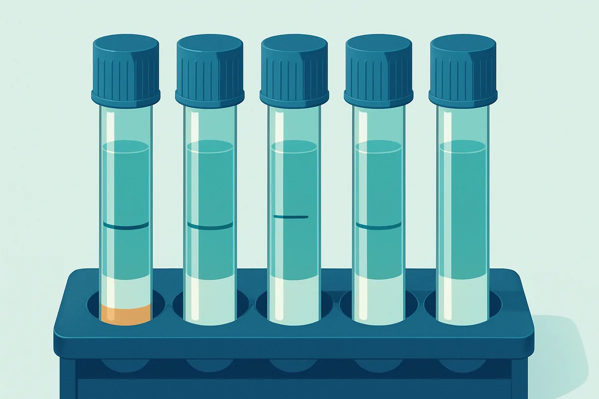
The procedure is simpler than its reputation. Blood is collected into citrate, which binds calcium and prevents clotting during transport. The tube is centrifuged to produce plasma, which is warmed. A reagent containing tissue factor, phospholipid and calcium is added, and a timer starts. The calcium reverses the citrate’s effect, the tissue factor triggers the cascade, and the instrument measures how long it takes for a clot to form.
Detecting the endpoint is where instruments differ. Optical instruments shine light through the sample and watch for the change in transmitted or scattered light as fibrin strands form and the plasma becomes turbid. Mechanical instruments detect the physical change directly, historically by monitoring a small steel ball whose movement is impeded as the sample gels.
The reagent is the other major variable, and it is the reason the raw result travels badly. Tissue factor preparations differ in origin, in whether the tissue factor is recombinant or extracted, in phospholipid composition, and in concentration. A plasma sample from a patient on warfarin will produce quite different clotting times with different reagents, and the more sensitive the reagent, the greater the prolongation for the same degree of anticoagulation. Each laboratory therefore establishes its own normal range for its own reagent and instrument combination, and a prothrombin time in seconds is meaningful only against that laboratory’s own reference values.
Why INR Exists and What ISI Corrects
The problem the INR solves is straightforward to state. In the era when oral anticoagulation became widespread, dosing was guided by prothrombin time ratios, and because reagents differed so much in sensitivity, the same patient could appear adequately anticoagulated in one hospital and dangerously over-anticoagulated in another. Patients travelled; results did not.
The solution was to define a reference: an international reference preparation of thromboplastin, against which every commercial reagent is compared. That comparison produces a single number for each reagent, the international sensitivity index, which expresses how responsive that reagent is relative to the reference. A reagent behaving identically to the reference has an index of one, and less sensitive reagents have higher values.
The INR is then calculated by taking the ratio of the patient’s prothrombin time to the mean normal prothrombin time for that laboratory, and raising that ratio to the power of the sensitivity index. Raising to a power is the key step: it is a non-linear correction that stretches or compresses the ratio scale so that reagents of different sensitivities converge on the same answer for the same plasma.
Two inputs must be right for this to work. The sensitivity index must be correct for the specific reagent lot on the specific instrument, since the index is not purely a property of the reagent and shifts with the detection system. And the mean normal prothrombin time must be determined locally from a group of healthy individuals rather than taken from a package insert, because it is a laboratory-specific quantity.
| Value | What it is | Comparable between laboratories | Main use |
|---|---|---|---|
| Prothrombin time in seconds | Raw clotting time with local reagent | No | Internal quality control, local reference range |
| Prothrombin ratio | Patient time divided by mean normal time | Only partly | Intermediate step in the INR calculation |
| International sensitivity index | Responsiveness of a reagent lot against a reference | Not applicable, it is a reagent property | Correcting the ratio to a common scale |
| INR | Ratio raised to the power of the index | Yes, within its validated setting | Monitoring vitamin K antagonist therapy |
The critical limitation is scope. The whole calibration system was established using plasma from patients stably anticoagulated with vitamin K antagonists, and the correction is validated for that population. Applied elsewhere it becomes approximate. In liver disease, the pattern of factor deficiencies differs from that produced by warfarin, and the correction that works for one does not transfer cleanly to the other, which is why prognostic scoring systems that incorporate the INR specify how it must be measured. In the first days of starting warfarin, before factors with longer half-lives have fallen, the INR rises largely because factor VII has dropped and overstates the degree of protection achieved.
aPTT and the Contact Activation Pathway
The activated partial thromboplastin time follows the same physical logic with a different trigger. Citrated plasma is incubated with a negatively charged activator, which mimics a foreign surface and switches on the contact factors, together with phospholipid. Calcium is then added and the clotting time measured. The word partial in the name refers to the absence of tissue factor: the reagent supplies phospholipid but not the protein component, so the tissue factor route is not available.
The aPTT serves three quite different purposes, and confusing them causes trouble. It screens for deficiencies of the contact and intrinsic factors, which is how haemophilia is typically first suspected. It detects certain inhibitors, notably the antiphospholipid antibodies that prolong the test by interfering with the phospholipid in the reagent while paradoxically associating with thrombosis rather than bleeding. And it is used to monitor unfractionated heparin therapy.
That last use has well-known weaknesses. Reagent sensitivity to heparin varies substantially, so each laboratory must establish its own therapeutic range by comparing aPTT results against a direct measure of heparin activity, and a range copied from elsewhere is meaningless. The relationship is also disturbed by the acute phase response: inflammation raises factor VIII and fibrinogen, both of which shorten the aPTT, so an acutely ill patient may show a deceptively short time despite adequate heparin. Many laboratories have moved towards measuring heparin activity directly for this reason.
Sample Volume, Citrate Ratio and Failed Tubes
No other routine test is as unforgiving about collection, and understanding why converts a set of rules into something obvious.
The citrate tube contains a fixed volume of liquid anticoagulant, calculated so that when filled to the line it produces a specific ratio of anticoagulant to blood, conventionally one part to nine. The citrate binds calcium in proportion to its amount. During the test, a fixed volume of calcium-containing reagent is added to reverse that binding. The entire timing therefore depends on the citrate concentration being what the method assumes.
An underfilled tube contains the full dose of citrate diluted into less blood, so the plasma carries excess citrate. The reagent’s calcium is partly consumed neutralising it, less remains to drive the reaction, and clotting takes longer. The result is a falsely prolonged PT and aPTT, and the effect is disproportionate: modest underfilling produces a modest error, but a substantially short tube can produce a dramatically wrong result. Because a raised INR looks like over-anticoagulation, the consequence is a warfarin dose reduced for no reason.
Haematocrit produces the same problem through a different route. The citrate volume assumes a normal proportion of plasma in the blood. In someone with a very high haematocrit there is less plasma per tube, so the same citrate is concentrated into a smaller plasma volume and the result is prolonged exactly as in underfilling. The correction is to use a tube with a reduced citrate volume, calculated for that patient, and laboratories apply this above a defined haematocrit threshold. Very low haematocrits create the mirror-image problem, though the effect is smaller.
Direct Oral Anticoagulants That Escape These Tests
Warfarin lowers the functional levels of several factors at once, which is exactly what the prothrombin time is good at detecting. The direct oral anticoagulants do something different: each inhibits a single enzyme, either thrombin or factor Xa, without changing how much of it is present. Screening tests respond to that inconsistently.
A direct thrombin inhibitor generally prolongs the aPTT, though the relationship flattens at higher concentrations so the test cannot quantify the effect. It affects the prothrombin time less. The direct factor Xa inhibitors tend to prolong the prothrombin time to a degree that depends heavily on which reagent the laboratory uses, with some reagents showing a clear response and others barely reacting at the same drug concentration.
The clinically important consequence is that a normal PT and aPTT do not exclude a therapeutic level of these drugs. A patient arriving with bleeding or requiring urgent surgery cannot be cleared on the basis of normal screening tests, and treating a normal result as evidence that no anticoagulant is on board is a genuine hazard.
Where the concentration must be known, specific assays exist. A dilute thrombin time or an ecarin-based assay quantifies direct thrombin inhibitors. An anti-Xa assay calibrated specifically against the relevant drug quantifies factor Xa inhibitors, and the calibration matters, since an anti-Xa assay calibrated for heparin will respond to a factor Xa inhibitor but will not report its concentration correctly.
Mixing Studies and Sorting Inhibitors From Deficiency
A prolonged clotting time has two broad explanations. Something necessary is missing, or something present is interfering. The mixing study distinguishes them using a single simple idea.
Patient plasma is mixed in equal parts with plasma from healthy donors and the test is repeated. Normal plasma contains a full complement of every factor, so mixing supplies roughly half-normal levels of everything. Because clotting times remain within the normal range down to factor levels well below half, a pure deficiency is corrected by this mixture and the clotting time returns to normal. If instead the prolongation is caused by an inhibitor, that inhibitor acts on the added normal factors as well, and the clotting time stays prolonged.
Antiphospholipid antibodies behave differently. They interfere with the phospholipid component of the reagent rather than attacking a specific factor, so they typically fail to correct immediately and do not worsen with incubation. Confirming them relies on demonstrating phospholipid dependence: repeating the test with an increased phospholipid concentration, which overwhelms the antibody and shortens the time.
A residual anticoagulant in the sample also fails to correct and is the most common reason for an uncorrected mixing study in practice, which is why the first question asked of any such result concerns what the patient is taking and how the sample was drawn.
The habit worth carrying away from all of this is to treat a coagulation result as the output of a defined laboratory procedure rather than a reading of the patient’s bleeding risk. Ask whether the tube was properly filled, since that single question explains a large share of unexpected results. Ask whether the INR is being used in the setting it was calibrated for. Ask what the patient is taking, because several widely used drugs are invisible to these tests and others distort them. And remember that a prolonged clotting time and a tendency to bleed are related but genuinely distinct findings, with well-established examples of each occurring without the other.
Frequently asked questions
Why did my INR change when nothing about my medication did?
Vitamin K intake is the usual explanation, since warfarin works by opposing vitamin K and the dietary supply varies day to day with green vegetables. Illness matters too: fever, infection and diarrhoea all shift the balance, and a great many drugs interact with warfarin’s metabolism or its protein binding, including some antibiotics, antifungals and over-the-counter preparations. Alcohol intake and changes in liver function contribute. A collection problem, most often an underfilled tube, should also be considered when a single result looks out of keeping with the pattern before and after it.
What does it mean if my aPTT is prolonged but I have never had a bleeding problem?
Several possibilities produce exactly that picture, and most are not bleeding disorders. A deficiency of factor XII prolongs the aPTT markedly and causes no bleeding at all, because that factor is not required for clotting in the body. An antiphospholipid antibody prolongs the test while being associated with clotting rather than bleeding. A collection artefact, particularly heparin contamination from a line or an underfilled tube, is common. Investigation usually begins with a repeat on a properly collected sample followed by a mixing study to establish whether something is missing or something is interfering.
Can the INR be used for anticoagulants other than warfarin?
Not reliably. The entire calibration system that converts a prothrombin time into an INR was established using plasma from patients on vitamin K antagonists, and its accuracy outside that setting is not assured. Direct oral anticoagulants affect the prothrombin time to a degree that varies with the reagent, so an INR calculated in a patient taking one carries no consistent meaning and cannot guide dosing. The INR is also used within specific prognostic scores for liver disease, where the measurement conditions are defined precisely because the value would otherwise not be comparable.
Why does the tube have to be filled exactly to the line?
Because the ratio between citrate anticoagulant and blood is part of the test method rather than an incidental detail. The tube holds a fixed amount of citrate designed to bind calcium in a full tube of blood, and the reagent added later contains a fixed amount of calcium to reverse that binding. An underfilled tube leaves excess citrate that consumes part of the reagent calcium, delaying clot formation and producing falsely prolonged times, which on an INR looks like over-anticoagulation. Laboratories reject underfilled coagulation tubes for this reason rather than out of procedural rigidity.
Do normal coagulation screens mean it is safe to have surgery?
Not by themselves. These tests were designed to investigate suspected abnormalities, and using them as a general pre-operative screen in people with no bleeding history has repeatedly been shown to identify little of value while generating false alarms, particularly through factor XII deficiency and incidental antiphospholipid antibodies. A careful bleeding history, including previous surgery, dental extractions, childbirth and family history, predicts surgical bleeding better. The tests also miss platelet function disorders entirely and will not reveal a direct oral anticoagulant reliably, so a normal screen does not establish that no anticoagulant is present.
This is education, not medical advice. Laboratory results only carry meaning alongside your symptoms, history and examination. Talk to a qualified clinician about your own results before changing anything about your care or supplements.



