

A blood culture is the test that decides whether an unwell patient has bacteria circulating in their bloodstream. It is slow by the standards of modern diagnostics, taking a day or more to give an answer, and it remains the reference against which everything faster is judged. When it grows a genuine pathogen, it usually changes treatment immediately.
The difficulty is that it grows things that are not pathogens surprisingly often. Blood is drawn through skin, skin is populated by bacteria that live in and around hair follicles and sweat glands, and no realistic disinfection procedure sterilises it completely. A few organisms carried in on the needle land in a warm bottle of rich broth and multiply happily for the next eighteen hours. The instrument then signals a positive result that looks, at the moment it appears, exactly like the real thing.
The consequences of that ambiguity are not trivial. A false positive commonly leads to additional antibiotics, extra days in hospital, repeat sampling and sometimes an intravenous line placed specifically to deliver treatment for an infection that was never there. Reducing contamination is therefore one of the highest-value quality projects any hospital laboratory runs, and interpreting the positives that do occur is a daily exercise in reading several weak clues together.
Key takeaways
- Blood culture bottles are nutrient broths with an internal sensor that detects the metabolic activity of growing bacteria.
- Volume of blood drawn is the single strongest determinant of whether an infection is detected, because bacteria are usually scarce.
- Skin flora contaminate a small but persistent proportion of sets, and the numbers depend heavily on collection technique.
- Species identity, how many bottles grew, and how quickly they signalled are the main clues separating contaminants from pathogens.
- Rapid identification from a positive bottle shortens the guessing period substantially but does not remove the need for interpretation.
How a Blood Culture Bottle Works
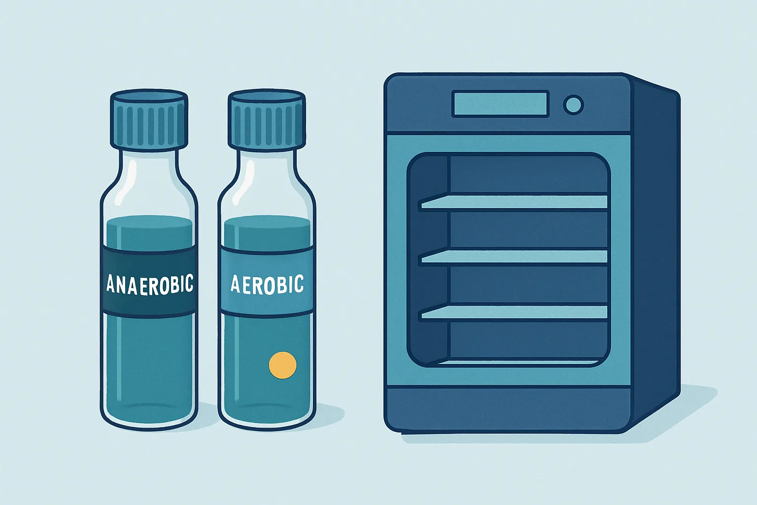
A blood culture bottle is a sealed glass or plastic vessel containing a nutrient broth designed to support the growth of a wide range of bacteria and yeasts from a very small starting number. Blood is injected through a rubber septum, and the bottle goes into an incubator held near body temperature.
Bottles come in pairs for a reason. One is formulated and gassed to support organisms that require oxygen, the other excludes oxygen to support those that cannot tolerate it. Many important bacteria grow in both, but a meaningful minority grow only in one, so a set of two covers a far wider range than either alone. Paediatric bottles exist as a separate format because the volume of blood that can safely be taken from a small child is much lower, and the broth is adjusted accordingly.
The broth contains more than food. Blood carries antibiotics if the patient has already been treated, and it carries the patient’s own antibacterial defences: complement proteins, antibodies, and white cells still capable of killing what they encounter. Culture media therefore include agents that neutralise or absorb antibiotics, most often resins or activated charcoal, and dilution of the blood into a much larger volume of broth reduces the effect of the patient’s own immune components. This is one reason overfilling a bottle is unhelpful even though underfilling is worse.
At the base sits a sensor. In the most widely used design, a small disc of material changes colour as carbon dioxide accumulates, because growing bacteria respire and produce it. An optical reader watches that disc every few minutes.
Why Volume Drawn Dominates Sensitivity
Bacteraemia in adults is often a low-density event. The concentration of organisms in blood can be low enough that a millilitre may contain none at all, which turns detection into a straightforward sampling problem: the more blood placed in the bottle, the higher the probability that at least one viable organism is captured.
This makes fill volume the dominant technical variable in blood culture sensitivity, ahead of media formulation, incubation protocol and instrument model. Underfilled bottles are the commonest reason a genuine infection goes undetected, and the effect is large rather than marginal. Laboratories that begin weighing bottles to monitor fill volume routinely discover that a substantial share of their bottles fall short of the recommended range, and that correcting it raises detection rates without any other change.
The same logic explains why more than one set is collected. Two sets drawn from separate venepuncture sites roughly double the blood volume examined, and they also create the comparison that makes contamination interpretable. A single set gives no way to ask whether an organism appeared in one draw or several.
Timing matters less than intuition suggests. There is a persistent belief that blood should be drawn at the peak of a fever, on the theory that organisms are most abundant then. In practice, bacteria typically enter the circulation before the temperature rises, since fever is the downstream response to that entry. Waiting for a spike delays treatment for no reliable gain. What does matter, decisively, is drawing cultures before antibiotics are given, because even a single dose can render subsequent bottles sterile while the infection continues.
Skin Antisepsis and Contamination Rates
The organisms that contaminate blood cultures are almost always the ones that live on skin: coagulase-negative staphylococci above all, along with corynebacteria, cutibacteria and various aerobic spore-formers. They reach the bottle either on the needle as it passes through the skin or from an inadequately disinfected bottle septum.
Skin cannot be sterilised. Bacteria occupy the deeper layers of hair follicles and glands where topical antiseptics do not reach, and a proportion are released when a needle passes through. Antisepsis reduces the surface population by a large factor and reduces the probability that any organism reaches the broth, but it cannot reach zero.
Technique details carry most of the variation between wards and between hospitals. Alcohol-based chlorhexidine preparations generally outperform aqueous iodine solutions, mainly because the alcohol acts quickly and the chlorhexidine keeps acting afterwards. Contact time is where practice most often fails: an antiseptic applied and immediately wiped or punctured has not had time to work, and the required drying period is skipped under pressure far more often than it is documented. The bottle tops themselves need disinfecting, since the plastic caps are not sterile underneath and a needle carries whatever sits on the septum straight into the broth.
Drawing blood cultures from an existing intravascular line rather than by fresh venepuncture raises contamination substantially, because the line’s hub and lumen are colonised even when the patient is not infected. Line draws have a legitimate role when line infection itself is the question, but as a routine convenience they degrade the test.
Dedicated phlebotomy teams consistently achieve lower contamination rates than ad hoc collection by whoever is available, which is less a comment on individual skill than on the value of doing the same procedure the same way many times a day.
Distinguishing Contaminants From True Pathogens
When a bottle signals positive, the laboratory and the clinical team assemble several independent clues. None is conclusive alone.
Species identity is the strongest. Some organisms are almost never contaminants: when they grow, an infection is presumed until proven otherwise. Others are overwhelmingly skin flora and only occasionally cause genuine disease, typically in people with prosthetic material or indwelling lines. The middle group is genuinely ambiguous and depends heavily on context.
| Organism group | Usual interpretation | Main caveat |
|---|---|---|
| Staphylococcus aureus | Nearly always significant | Requires source hunting even when the patient looks well |
| Streptococcus pneumoniae | Nearly always significant | Rarely a contaminant at all |
| Escherichia coli and related bowel organisms | Nearly always significant | Very unlikely to come from skin |
| Candida species | Nearly always significant | Prompts assessment for line infection and spread to the eye |
| Coagulase-negative staphylococci | Usually contamination | Genuine when prosthetic material or a line is present |
| Corynebacteria and cutibacteria | Usually contamination | Slow growers, occasionally real in device infection |
| Bacillus species other than anthracis | Usually contamination | Occasionally real in injecting drug use |
The second clue is how many bottles grew. An organism recovered from two separate sets, drawn at different sites, is far more likely to have come from the blood than from skin, since contamination of two independent punctures with the same organism is improbable. A single bottle of four growing a skin organism points strongly the other way.
The third is clinical coherence. A patient with a fever, a raised inflammatory response and no other explanation is a different proposition from a well patient whose culture was drawn as a precaution. And in ambiguous cases, whether the same organism reappears on a repeat set often settles the matter.
Time to Positivity as a Clue
Automated systems record the exact hour a bottle signals, and that number carries information that is easy to overlook.
Time to positivity reflects roughly how many organisms were present at the start and how fast that species grows. A bottle containing many organisms crosses the detection threshold sooner than one seeded with a handful. Genuine bloodstream infections with rapidly growing organisms typically signal within the first day, often within half of it. Contaminants introduced as a few skin bacteria tend to take longer, and the slow-growing skin genera can take two days or more.
The clue is soft rather than decisive, because a real infection caught early or treated partially can also grow slowly. It is most useful in combination: a skin organism from one bottle only, signalling late, is very likely contamination, while the same organism from two sets signalling early deserves to be taken seriously.
There is a second, more specific use. When cultures are drawn simultaneously from a central line and from a peripheral vein, and the line bottle signals substantially earlier than the peripheral one, that difference suggests the organism concentration was higher inside the line, which points to the line itself as the source. This differential timing has become a standard part of assessing catheter-related infection, and it requires only that both sets are drawn at the same time and clearly labelled.
Automated Detection and Flagging Systems
Modern blood culture instruments are essentially large incubators full of optical readers. Each bottle position is monitored every ten to fifteen minutes, and the reading is not a simple threshold but a trend.
The reason for watching the trend rather than an absolute value is that blood itself varies. A bottle from a patient with a high white cell count starts with more background metabolic activity, and a fixed threshold would flag it spuriously. Detection algorithms therefore look for the characteristic acceleration of an exponentially growing population: a rate of change, and a change in that rate, rather than a level.
When the algorithm fires, an alarm sounds and the bottle is removed for immediate examination. The first step is a Gram stain, which within minutes reports whether organisms are present and what broad shape and staining category they fall into. That single piece of information is often enough to change antibiotic therapy on the spot, hours before an identification is available.
Instruments also produce false negatives and false positives of their own. Some organisms grow without generating much carbon dioxide, and a few clinically important ones are recovered only when bottles are examined at the end of the incubation period despite never signalling. In the other direction, a bottle overfilled with blood from a patient with very high white cell activity can trigger an alarm with no organisms visible on the stain.
Standard incubation runs for five days in most laboratories. Extending it beyond that was once routine for slow-growing organisms associated with heart valve infection, but improved media have made prolonged incubation largely unnecessary for that purpose, and the additional time mostly yields contaminants.
Rapid Identification From Positive Bottles
Once a bottle is positive, it contains a large, nearly pure population of the organism, which makes it excellent material for rapid analysis. This has been the main area of progress in the field.
Mass spectrometry has become the standard identification method in many laboratories. Proteins from the organism are ionised and their mass pattern compared against a reference library, giving a species name in minutes rather than the day required by traditional biochemical panels. Applied directly to material from a positive bottle after a short preparation step, it can deliver an identification within an hour or two of the alarm.
Molecular panels go further by detecting a defined set of common organisms and selected resistance genes directly from the positive broth. Knowing within an hour that an organism carries a resistance determinant, or that the growth is a species requiring specific treatment, can shorten the period of broad empirical therapy considerably.
These methods identify. They do not, by themselves, measure susceptibility across the full range of antibiotics, which still generally requires growth in the presence of drug and therefore additional time, although shortened phenotypic methods are narrowing that gap.
Crucially, faster identification also makes contamination easier to handle. Learning within two hours that the organism is a common skin commensal, rather than the next morning, avoids a night of unnecessary treatment. Speed helps most when it is paired with the interpretive framework: what grew, from how many bottles, how fast, and in a patient who looks how.
Frequently asked questions
Why were two or more sets taken from different arms?
Two separate venepunctures serve two purposes at once. They roughly double the total volume of blood examined, which directly raises the chance of capturing organisms present in low numbers. They also create the comparison that makes a positive interpretable, because an organism appearing in both independent draws is very unlikely to have come from skin twice, whereas growth in one bottle of four points towards contamination. Sampling two sites is not duplication for safety, it is part of how the test is designed to be read.
Does a negative blood culture rule out infection?
No. It substantially lowers the probability of a bloodstream infection with an organism that grows in standard media, but several situations produce negative results despite genuine infection. Antibiotics given before the draw are the commonest, and even one dose can sterilise the sample. Some organisms grow poorly or not at all in routine bottles and require specific media or molecular testing. Infection confined to a tissue site without spilling into the blood also gives negative cultures, and inadequate fill volume remains a frequent technical cause.
What does it mean when only one bottle out of four grows something?
It raises the probability of contamination considerably, particularly if the organism is a common skin inhabitant and the bottle signalled late. It does not settle the question, because low-level bacteraemia can genuinely seed only one bottle, especially when fill volumes were uneven. Interpretation weighs the species, the patient’s clinical state, whether any prosthetic material or intravascular line is present, and what a repeat set shows. A single positive bottle growing an organism that is almost never a contaminant is treated as real.
Why does it take so long to get a final result?
The test depends on multiplying a possibly single organism into a population large enough to detect, which is biology operating at its own speed. Most clinically important bacteria signal within a day, but the bottles are held for several days to catch slower growers, so a genuinely negative report cannot be issued until that period ends. After a positive, identification is now often quick, while susceptibility testing usually requires further growth in the presence of antibiotics before reliable results can be reported.
Can contamination be prevented entirely?
Not entirely, because bacteria persist in skin structures that surface antiseptics cannot reach. It can be reduced a long way. The measures that produce most of the improvement are consistent use of an alcohol-based antiseptic with a full drying time, disinfection of the bottle tops, fresh venepuncture rather than drawing from an existing line, correct fill volume, and having the procedure performed by people who do it often. Hospitals that monitor their contamination rate and feed the number back to the teams collecting samples reliably see it fall.
For anyone reading their own result, the useful questions are which organism grew, how many bottles it grew in, and whether the treating team considered it a contaminant. A positive blood culture is a piece of evidence rather than a verdict, and the interpretation that surrounds it, drawing on species, number of positive sets, timing and clinical picture, is where the actual diagnosis is made.
This is education, not medical advice. Laboratory results only carry meaning alongside your symptoms, history and examination. Talk to a qualified clinician about your own results before changing anything about your care or supplements.



