

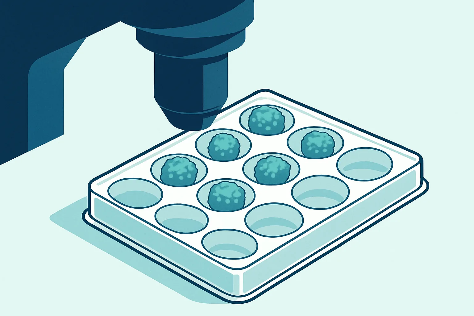
Reports about organoids tend to reach for the phrase “mini-organ”, which is a good headline and a poor description. What sits in the well is a few hundred micrometres across, contains several cell types arranged in something resembling the layered structure of a real tissue, and has no blood supply, no immune system and no nerves.
That combination is genuinely useful and genuinely limited, and the two are inseparable. Organoids reproduce the local architecture of a tissue, meaning which cell types form and where they sit relative to one another, far better than cells grown flat on plastic. They cannot reproduce anything that depends on being connected to the rest of a body, because they are not.
The interesting question is not whether organoids are impressive. It is which specific biological questions their structure allows them to answer, and which questions their missing components put permanently out of reach unless something is added back. This piece tries to draw that line clearly.
Key takeaways
- An organoid is a self-organising three-dimensional culture that reproduces some of a tissue’s architecture and cell type composition.
- Cell polarity, layered structure, tissue-specific function and differentiation are reproduced well.
- Blood vessels, immune cells and nerves are absent, which sets a size limit and removes whole categories of biology.
- Batch-to-batch variability is the main practical barrier to using organoids in large-scale screening.
- They complement animal models rather than replacing them, particularly where whole-body responses matter.
What an Organoid Physically Is
An organoid is a three-dimensional culture, grown from stem cells or tissue-derived progenitors, that organises itself into structures resembling the tissue of origin without being told how. The self-organisation is the defining feature and the surprising one.
Nobody arranges the cells. Given the right starting population and the right chemical environment, the cells arrange themselves, and they do so because the instructions are already inside them. Cells signal to their neighbours, respond to those signals by adopting particular fates, position themselves according to adhesion preferences and mechanical forces, and the result is a structure with recognisable geometry. Intestinal organoids form crypt-like invaginations with proliferating cells at their base and differentiated cells further out, which is the arrangement of a real intestinal lining. Kidney organoids form structures resembling nephron segments. Brain organoids develop layered zones reminiscent of the developing cortex.
Physically the culture requires a scaffold. Cells are usually embedded in a hydrogel matrix that provides mechanical support and the adhesion signals a basement membrane would normally supply. Commonly used matrices are derived from tumour cell lines and are chemically complex and somewhat variable between batches, which is a recognised weakness driving work on chemically defined synthetic alternatives.
Size is limited to a few hundred micrometres to a couple of millimetres, and the reason is oxygen and nutrient diffusion. Without vessels, everything reaches the interior by diffusion alone, and beyond a certain radius the centre becomes hypoxic and dies. That necrotic core is not an occasional artefact; it is the predictable consequence of a geometry that has no delivery system, and it sets a hard ceiling on how large an organoid can become while remaining viable throughout.
Stem Cell Sources and Differentiation Cues
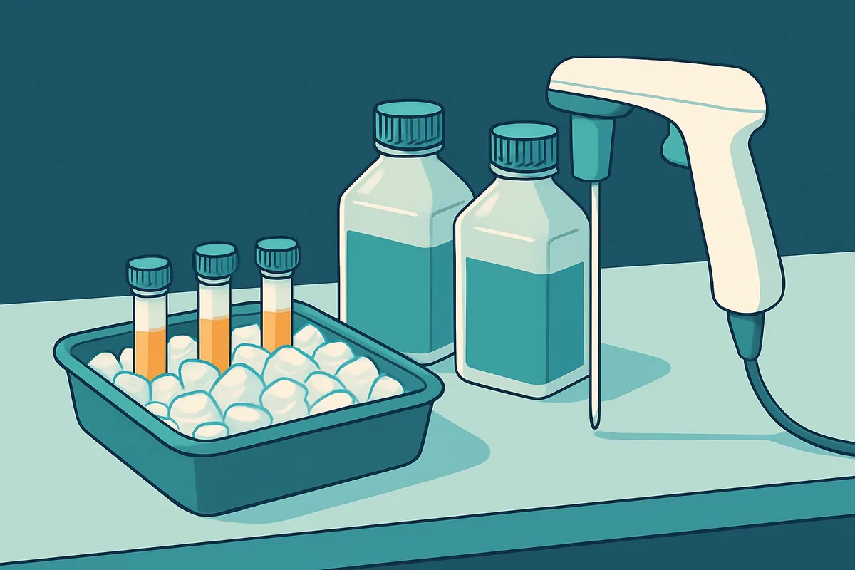
Two broadly different starting materials produce organoids with different properties, and knowing which was used tells you a great deal about what a given model can do.
Pluripotent stem cells, either embryonic or induced from adult cells, can in principle form any tissue. Making an organoid from them means recapitulating development: applying sequences of signalling molecules that push cells through the same decision points an embryo would, from germ layer specification onward. This is powerful because it grants access to tissues with no accessible adult stem cell population, brain being the clearest example. It also means the resulting tissue is developmentally immature, resembling a fetal stage rather than an adult one, and maturation remains one of the field’s central unsolved problems.
Adult tissue-derived stem cells offer the opposite trade. Taken from a biopsy of intestine, liver, pancreas, lung or several other tissues, they produce organoids that are already adult in character and match the donor genetically. They can only make the tissue they came from, which limits scope but removes the maturation problem entirely, and they are the basis of patient-derived organoids used to model individual disease.
The differentiation cues themselves are the signalling pathways that pattern the embryo, applied in sequence with careful timing. Concentration and duration both matter, because the same pathway can specify different fates depending on how strongly and how long it is activated. Protocols are therefore long, multi-step and sensitive, and small deviations propagate. This sensitivity is a major source of the variability discussed later.
| Source | Tissues accessible | Maturity | Main use |
|---|---|---|---|
| Embryonic stem cells | Broadly any | Fetal-like | Development, tissue types with no adult stem cells |
| Induced pluripotent cells | Broadly any | Fetal-like | Patient-specific genetics, disease modelling |
| Adult tissue stem cells | Only the tissue of origin | Adult | Patient-derived models, epithelial disease |
| Tumour tissue | The tumour’s own lineage | Matches tumour | Cancer drug response, biobanking |
Structures These Models Reproduce Well
What organoids do better than flat culture is anything that depends on cells knowing which way is up.
Epithelial polarity is the clearest example. Real epithelia have an apical surface facing a lumen and a basolateral surface facing the underlying tissue, and the two carry entirely different transporters, receptors and junctions. A cell grown flat on plastic has no meaningful lumen and its polarity is a poor approximation. In an organoid, epithelial cells form a genuine lumen with correctly oriented apical surfaces, which matters enormously for anything involving secretion, absorption or barrier function. Studies of ion transport, drug absorption across an epithelium, and pathogens that infect from the apical side all become possible in a way they simply were not before.
Cell type composition is the second strength. A tissue is a mixture, and the proportions matter. Intestinal organoids generate absorptive cells, mucus-secreting cells, hormone-producing cells and the stem cells that maintain them, in roughly appropriate ratios and positions. That heterogeneity supports questions about how cell types interact locally, which a pure population cannot address.
Tissue-specific function follows from the structure. Liver organoids perform metabolic functions that flat hepatocyte cultures lose rapidly. Intestinal organoids transport ions and respond to secretory stimuli. Kidney organoids show filtration-related structures. Function tends to be present but reduced relative to the real tissue, which is a consistent pattern across the field and an important caveat.
Developmental processes are the fourth strength, particularly for pluripotent-derived models. Because the organoid recapitulates development to reach its final state, it can be observed while doing so. This has been especially valuable for human brain development, where the equivalent tissue is otherwise inaccessible and where mouse development differs from human in ways that matter.
Missing Vasculature and Immune Components
The absences are structural, not incidental, and they define the boundary of what organoids can address.
Vasculature is the most consequential. Blood vessels do more than deliver oxygen. They establish gradients of nutrients and signalling molecules across a tissue, remove waste, provide the endothelial signals that many tissues need for maturation, and serve as the route by which circulating drugs and cells arrive. Without them an organoid is limited in size, develops a necrotic core if it exceeds that limit, lacks physiological gradients, and cannot model anything involving delivery through the circulation. Considerable effort has gone into vascularising organoids, either by co-culturing endothelial cells or by transplanting organoids into an animal where host vessels grow in. Both work partially and neither is routine.
Immune components are absent for a similar reason: they are recruited from elsewhere in the body. This removes inflammation, immune surveillance, immune-mediated tissue damage and the resident immune populations that many tissues normally contain. For inflammatory bowel disease, autoimmune conditions, infection biology and cancer immunotherapy this is not a detail but the central mechanism. Co-culture with immune cells is an active area and adds one or two cell types rather than an immune system.
Nerves are absent from most organoids too, which matters for gut motility, secretion, and any tissue where neural input shapes function.
Mechanical and systemic context is the fourth gap and is easily overlooked. Real tissues experience flow, stretch, peristalsis and breathing motion, and cells respond to mechanical force by changing gene expression. A static organoid in a gel experiences none of it. Organ-on-chip approaches address precisely this by adding controlled flow and cyclic strain, and combining them with organoid biology is one of the more promising current directions.
Finally, there is no other organ. A drug metabolised by the liver into a toxic product will not show that toxicity in a kidney organoid, because there is no liver upstream.
Batch Variability Between Organoids
The problem that most constrains practical use is that organoids differ from one another, sometimes substantially, even within a single experiment.
The cause follows from self-organisation. Because the structure emerges from cells interacting rather than from an imposed plan, small differences in starting conditions amplify. The number of cells in the initial aggregate, its exact shape, its position in the gel, local differences in matrix stiffness, and slight variations in when signalling molecules were added all influence the outcome. Two organoids from the same starting culture can end up different sizes, with different proportions of cell types and different degrees of organisation.
The matrix contributes its own variation, since tumour-derived hydrogels vary between production lots in composition and mechanical properties. Cell line background contributes more, as different induced pluripotent lines differentiate with different efficiencies, so comparing a patient line to a control line risks measuring the lines rather than the disease.
This has direct consequences. Statistical power falls, so more organoids are needed to detect a given effect. Modest differences between conditions can be swamped by variability between organoids. Comparisons across laboratories become difficult, and reproducibility suffers.
Several approaches reduce it. Controlling starting aggregate size using microwells produces more uniform structures. Chemically defined matrices remove one variable. Bioreactors improve consistency of nutrient and oxygen exposure. Isogenic controls, made by editing a mutation into or out of a single cell line, eliminate genetic background as a confounder and are now regarded as close to essential for disease modelling. Automated imaging with quantitative morphology allows organoids to be characterised and filtered rather than assumed equivalent.
None of these eliminates the problem, and honest reporting of variability, including how organoids were selected for analysis, matters as much as reducing it.
Applications in Drug Screening
Despite the variability, organoids have found real footing in drug work, and it is worth being specific about where.
Patient-derived cancer organoids are the most established application. Tumour tissue from a patient is grown as organoids, exposed to candidate drugs, and the response measured. These models retain much of the original tumour’s genetic character and cellular heterogeneity, which cancer cell lines, adapted over decades in flat culture, largely do not. Biobanks of tumour organoids paired with clinical data have become a substantial resource, and studies comparing organoid drug response to the patient’s actual clinical response have shown encouraging concordance for some tumour types and treatments, though this remains an area of active evaluation rather than settled practice.
Toxicity screening is the second area. Liver and kidney organoids retain more tissue-specific function than the cell lines conventionally used, so they detect some toxic effects that flat cultures miss. The caution is that toxicity often depends on metabolism in one organ affecting another, which a single organoid cannot capture.
Rare disease modelling is the third and is where organoids arguably have the least competition. For a condition affecting very few people, generating induced pluripotent cells from a patient and differentiating them into the affected tissue may be the only route to a human model at all.
Infection biology has been productive, because correctly polarised epithelium with a genuine apical surface is what many pathogens require to infect in a realistic way.
The general position is that organoids are strongest for mechanistic questions and for patient-specific prediction, and weakest for large-scale primary screening where throughput and consistency dominate. They are widely used as a secondary filter: cell lines narrow a large library, organoids test the survivors under more realistic conditions.
Where Animal Models Still Do More
Framing organoids as replacements for animal experiments is common in coverage and is not what the science supports. The two answer different questions.
An animal has everything an organoid lacks. It has a circulation, so a drug administered to it distributes, is metabolised, is excreted, and reaches tissues at concentrations determined by real pharmacokinetics. It has an immune system that responds to what is happening. It has multiple organs that influence each other, so liver-mediated toxicity in the kidney appears without anyone designing for it. It has behaviour, which is the only readout for a great deal of neuroscience, pain research and psychiatry. It has a lifespan, so chronic exposure and progressive disease can be observed over time.
Where organoids genuinely do more is human specificity, developmental accessibility, mechanistic dissection and scale. Human biology differs from mouse biology in ways that have caused real translational failures, and a human organoid speaks directly to human cells. Human developmental processes can be watched in a dish. Genetic manipulation and imaging are far easier in a controlled culture than in a living animal. And an experiment can be run across many conditions at once.
The productive arrangement uses both. Organoids identify mechanisms and generate candidates in human cells, animals test whether those findings hold in a whole organism with circulation and immunity, and organoids again help interpret what the animal showed. This also serves the reduction and refinement principles that govern animal research, because better human models mean fewer animals used on candidates that were never going to work.
What organoids will not do soon is replace whole-organism testing for questions that are inherently about the whole organism. Progress on vascularisation, immune co-culture, chip-based multi-organ systems and maturation is steady and real, and each closes part of the gap. None has closed it.
Frequently asked questions
Are organoids the same thing as lab-grown organs?
No, and the distinction matters. An organoid is a small, structurally simplified model of part of a tissue, typically under a few millimetres, lacking blood vessels, nerves and immune cells. It is a research tool. Growing a transplantable organ requires a vascular network that can be connected to a recipient’s circulation, appropriate size and mechanical strength, and full functional maturity, none of which current organoids possess. Work toward transplantable tissue exists but is a distinct and much earlier-stage effort.
Do brain organoids have any form of consciousness?
The scientific consensus is clearly no. Brain organoids reproduce some aspects of cortical development and generate spontaneous electrical activity, which is a property of neural tissue rather than evidence of experience. They lack sensory input, motor output, the connectivity between brain regions that integrated function requires, and anything resembling whole-brain organisation. Researchers and ethicists nonetheless discuss the question actively, because the appropriate time to establish boundaries is before rather than after a threshold is approached.
Why do organoids stay so small?
Because they have no blood vessels, so oxygen and nutrients reach the interior only by diffusion. Diffusion becomes inadequate beyond a few hundred micrometres, and past that the centre becomes hypoxic and forms a necrotic core. This is a physical constraint rather than a technical shortcoming, which is why vascularisation is such a prominent research goal, and why transplantation into a host animal, where blood vessels grow in, allows organoids to reach larger sizes.
Can an organoid predict how a specific patient will respond to a drug?
For some cancers this has shown genuine promise, with tumour organoid responses corresponding reasonably well to clinical outcomes in several studies. It is not yet a routine clinical tool. The limitations are that organoids lack the immune component central to many modern cancer therapies, that not all tumours grow reliably as organoids, that turnaround time may exceed clinical need, and that variability complicates interpretation of a single patient’s result.
Will organoids eliminate the need for animal testing?
They will reduce it in specific areas and will not eliminate it. Organoids cannot model drug distribution through a circulation, whole-body metabolism, immune responses, interactions between organs, behaviour, or long-term disease progression. Those are precisely the questions much animal research exists to answer. The realistic trajectory is that better human models cut the number of animals used on questions that a dish can answer, while whole-organism questions continue to require whole organisms until something can substitute for a body.
The useful test when reading about a new organoid result is to ask what the finding depended on. If it depended on epithelial architecture, cell type composition, human-specific genetics or a developmental process, the model was well suited and the result carries weight. If it depended on delivery through blood, on an immune response, or on one organ affecting another, ask what was added back and how convincingly. That single question separates the claims that will hold from the ones that will need a mouse to check.
This is education, not medical advice. Laboratory results only carry meaning alongside your symptoms, history and examination. Talk to a qualified clinician about your own results before changing anything about your care or supplements.



