

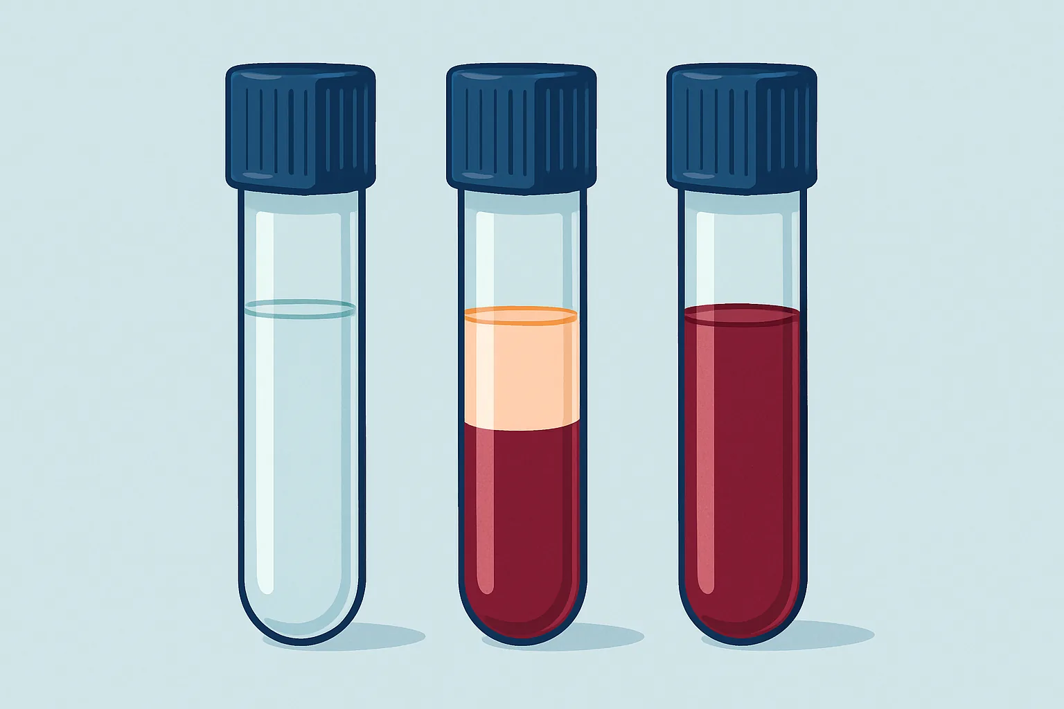
Being told a blood sample was unusable is irritating in a specific way. Nothing appeared to go wrong. The needle went in, the tubes filled, the plaster went on, and days later a message arrives asking you to come back and do it again. The explanation offered is usually one word, and it is usually haemolysis.
Haemolysis means that red blood cells have ruptured and released their contents into the surrounding plasma or serum. It is the most frequent reason samples are rejected in clinical laboratories by a wide margin, and it accounts for a large share of all pre-analytical error. Crucially, it is not a finding about the patient in the vast majority of cases. It is damage that happened during collection, handling or transport.
What makes haemolysis interesting rather than merely annoying is how selective its effects are. It does not degrade a sample uniformly: it devastates a small number of results, subtly distorts a few more, and leaves the majority untouched. Understanding which is which explains why a laboratory sometimes reports half a panel and suppresses the rest.
Key takeaways
- Haemolysis releases the contents of red cells into plasma, and those contents are wildly different in concentration from plasma itself.
- Potassium is affected most severely because its concentration inside red cells is many times higher than outside.
- Released haemoglobin also interferes optically and chemically, which affects assays with no connection to red cell contents.
- Most haemolysis is caused at the draw or in transport, not by anything happening inside the patient.
- Laboratories now measure haemolysis instrumentally and suppress affected results rather than rejecting whole samples.
What Ruptured Red Cells Release Into Plasma
A red blood cell is a bag of concentrated haemoglobin held apart from the plasma by a membrane maintaining steep gradients across itself. When that membrane breaks, everything inside spills into a compartment where those substances are normally present at very different concentrations, and the size of the resulting error depends on how steep the gradient was.
Haemoglobin is the most abundant release and the most visible, giving haemolysed plasma its pink or red tint at an intensity roughly proportional to the amount released. Because red cells are so densely packed with it, even a small proportion rupturing produces a visible change.
Potassium follows, and it is the most clinically dangerous release. Lactate dehydrogenase, an enzyme abundant in red cells, is released in quantities that raise plasma levels dramatically. Aspartate aminotransferase, one of the two enzymes used to assess liver function, is similarly concentrated inside red cells. Magnesium and phosphate are both higher inside cells than outside, so both rise.
Alongside the additions come distortions. Free haemoglobin absorbs light strongly across parts of the visible spectrum, and a great many assays work by measuring absorbance, so a coloured sample interferes with the measurement itself, independently of what was released. Free haemoglobin also participates in the chemistry of some assays and binds to certain reagents.
One final consideration matters clinically. Haemolysis can occasionally be real, meaning cells are being destroyed inside the patient rather than in the tube. In vivo haemolysis is uncommon and looks different: it is accompanied by a falling haemoglobin, a rising reticulocyte count, raised bilirubin, low haptoglobin and abnormalities on a blood film. Where those features are absent and only the tube is pink, the damage happened outside the patient.
Why Potassium Is the First Casualty
Potassium is the analyte most sensitive to haemolysis, and the reason is a single, very large concentration gradient.
Every cell maintains a high internal potassium and low internal sodium concentration, using the sodium-potassium pump to do so against the gradient. Inside a red cell, potassium sits roughly twenty-five to thirty times higher than in surrounding plasma. Sodium runs the other way, but plasma sodium is so high in absolute terms that dilution by cell contents barely moves it.
That asymmetry is what makes potassium so vulnerable. Because plasma potassium is normally a small number and the intracellular reservoir an enormous one, rupturing even a small percentage of red cells raises the measured value substantially. Well before the plasma looks obviously red, the potassium result can have moved from mid-range to a level that would prompt urgent action.
That is why a spuriously raised potassium is one of the more dangerous laboratory errors. Genuine severe hyperkalaemia is an emergency because it disturbs cardiac conduction, and it triggers treatment that lowers potassium. Applying that treatment to a patient whose potassium is normal causes harm. Laboratories therefore treat unexpected raised potassium with suspicion, and one calling to ask whether the draw was difficult is doing exactly the right thing.
The same gradient logic creates a related artefact. In samples with very high white cell or platelet counts, potassium can leak from those cells during clotting even without visible haemolysis. Cooling a sample before separation also slows the membrane pumps and lets potassium drift outwards, which is why refrigerating whole blood before spinning is discouraged.
Analytes That Survive Mild Haemolysis
Not every result is compromised, and this is where the selectivity becomes useful.
Analytes are largely unaffected when three conditions hold: the substance is not concentrated inside red cells, the assay does not measure absorbance where haemoglobin interferes, and haemoglobin does not react with the assay chemistry.
| Analyte | Effect of haemolysis | Underlying reason |
|---|---|---|
| Potassium | Large false increase | Concentration inside red cells is many times plasma level |
| Lactate dehydrogenase | Large false increase | Enzyme highly abundant in red cells |
| Aspartate aminotransferase | Moderate to large false increase | Present at high concentration inside red cells |
| Magnesium, phosphate | Moderate false increase | Higher inside cells than in plasma |
| Bilirubin | Variable, often falsely decreased | Optical and chemical interference from haemoglobin |
| Troponin | Assay-dependent, may fall or rise | Interference varies by manufacturer’s method |
| Sodium, chloride | Minimal change | Plasma concentration already high; cell content similar or lower |
| Urea, creatinine | Minimal change | Not concentrated inside red cells |
| Alanine aminotransferase | Small increase only | Much less abundant in red cells than AST |
| Most hormones and proteins | Usually minimal at mild grades | Not stored in red cells; assays less optically sensitive |
Two patterns in that table repay attention. The first is that AST rises far more than ALT with haemolysis, so a haemolysed sample can produce an AST-to-ALT ratio that mimics a specific pattern of liver disease. A laboratory that reports both without flagging haemolysis can send a clinician down an entirely unnecessary path.
The second is bilirubin’s behaviour, which moves opposite to most affected analytes. Haemoglobin interferes with the colour development used in bilirubin assays, typically producing a falsely low result. Intuition suggests bursting red cells should raise bilirubin, since bilirubin comes from haemoglobin breakdown, but that breakdown takes hours in the body and does not occur meaningfully in a tube.
Full blood counts occupy a special position. Haemolysis in the EDTA tube destroys the very cells being counted, so the red cell count and haematocrit fall while the measured haemoglobin, determined chemically after deliberately lysing all the cells, stays roughly the same. The indices become internally inconsistent, and a badly haemolysed count cannot be salvaged.
How Labs Grade Haemolysis Indices
For most of the history of laboratory medicine, haemolysis was assessed by eye. A technologist judged the separated sample clear, slightly pink, pink or red. That approach is subjective, varies with lighting and observer, and misses low-level haemolysis entirely.
Modern analysers measure it. A small aliquot is scanned at several wavelengths before analysis, and the absorbance pattern yields three separate indices: one for haemolysis, one for icterus and one for lipaemia. The haemolysis index derives from absorbance at wavelengths where free haemoglobin absorbs strongly, corrected for the other two interferents. The output is a number, usually a semi-quantitative scale or an estimated free haemoglobin concentration.
That number drives an automated decision. Each assay has a defined haemolysis threshold, established by the manufacturer through interference studies in which increasing amounts of haemolysate are added and the deviation from the true value measured. The threshold sits where interference exceeds acceptable analytical error. When a sample’s index exceeds an assay’s threshold, that result is suppressed or commented, while assays with higher thresholds are released normally.
This is why a laboratory can report most of a panel and withhold the potassium and LDH. It is not inconsistency; it is the same measured index compared against different tolerances.
Two limitations are worth knowing. Thresholds differ between manufacturers and platforms, so the same sample can be reportable in one laboratory and suppressed in another. And the index measures free haemoglobin, so it cannot distinguish haemolysis in the tube from haemolysis in the patient; that requires clinical information the analyser does not have.
Draw Technique Causes Versus Transport Causes
Most haemolysis is created at the moment of collection, and the causes are mechanical and identifiable.
Difficult venepuncture leads the list. Probing with the needle damages tissue and cells directly. A vein that collapses under the vacuum of an evacuated tube produces turbulent, intermittent flow, and turbulence shears red cells. Drawing through a fragile vein with a needle sized for a larger one has the same effect, as does drawing from an area with an existing haematoma.
Transfer technique matters as much as the puncture. Blood drawn into a syringe and then forced through a needle into a tube is being pushed through a narrow opening under pressure, which is an efficient way to rupture cells. Proper transfer devices let the tube’s own vacuum draw the blood in gently.
Alcohol that has not dried before puncture lyses cells at the entry site. Vigorous shaking to mix additive tubes, rather than gentle inversion, damages cells mechanically. Underfilled tubes leave excess additive relative to blood, and some additives are damaging at high relative concentration.
Transport and handling add a second set of causes. Pneumatic tube systems accelerate and decelerate carriers sharply, and a poorly padded carrier or an aggressive route can generate enough force to rupture cells; laboratories investigating unexplained clusters have repeatedly traced them to a particular route or carrier design. Prolonged delay before separation allows cells to fragment on their own. Freezing whole blood destroys cells completely, which is why samples must be separated before any freezing step.
Centrifugation errors form a third group: spinning too fast, spinning a serum tube before the clot has fully formed, or re-spinning a tube after separation can all rupture cells at the interface.
The pattern that distinguishes the causes is usually clustering. Haemolysis concentrated in samples from one collection area points at technique or equipment there. Haemolysis distributed across all sources but arriving through one transport route points at transport. Haemolysis in a single sample from an otherwise clean stream usually means a difficult draw.
Icterus and Lipaemia as Related Interferences
Haemolysis is one of three interferences measured together, and the other two follow similar logic with different chemistry.
Icterus is a yellow or brown discolouration caused by raised bilirubin. Unlike haemolysis, it is almost always a genuine patient finding rather than a handling artefact, reflecting liver disease, biliary obstruction or increased red cell breakdown. It interferes optically, absorbing strongly in the blue region, and chemically with certain assays, notably some creatinine methods. Because the cause is real, an icteric sample cannot be improved by redrawing, and laboratories must use alternative methods or report with a comment.
Lipaemia is a milky turbidity caused by suspended lipoprotein particles, principally chylomicrons carrying triglycerides. It arises from recent fat intake, intravenous lipid infusions, or disorders of lipid metabolism. Its interference is mostly physical: the particles scatter light, corrupting any absorbance-based measurement. It also creates a volume displacement effect, since lipid occupies space that water would otherwise occupy, which can falsely lower the measured concentration of substances dissolved in the aqueous phase.
Lipaemia is the one of the three that can often be corrected. Ultracentrifugation separates the lipid layer, and lipid-clearing reagents allow many assays to be salvaged. A fasting redraw usually resolves diet-related lipaemia entirely, which is why the fasting requirement exists for lipid panels.
Grouping the three together is analytically sensible because they interfere with the same measurement principle and their spectral contributions overlap. Separating them requires measurement at multiple wavelengths, which is exactly what the serum indices do.
Deciding Between Reporting and Redrawing
The decision a laboratory faces with a haemolysed sample is a balance between reporting something potentially wrong and delaying a result that may be needed urgently.
The default is straightforward. Where an analyte’s interference threshold is exceeded, the result is suppressed and a redraw requested. Where it is not, the result is released, often with a comment noting the presence of haemolysis so that anyone interpreting it knows the sample was not pristine.
Complications arise at the margins. A sample from a patient who is extremely difficult to bleed, or a paediatric sample obtained with considerable effort, carries a real cost to redrawing. So does one that cannot be recollected in the same clinical state, such as a sample drawn at a specific point in a drug dosing cycle. In these situations a laboratory may release a result with a strong caveat and an explicit statement of the direction the interference would push it. That is defensible when the direction is known and the clinical question is not sensitive to it, and indefensible when a decision depends on the exact value.
Timing changes the calculation too. In an emergency, an hour’s delay for a redraw may matter more than moderate uncertainty, and a blood gas analyser can often deliver a potassium quickly on a fresh sample drawn with minimal manipulation.
For anyone on the receiving end of a redraw request, the practical points are limited but real. Being well hydrated makes veins easier to access, reducing the probing and vein collapse that cause most haemolysis. Mentioning previous difficult draws lets the phlebotomist plan. Beyond that, the causes sit on the collection side rather than the patient side.
The larger point is that a rejected sample represents a laboratory doing its job rather than failing at it. The alternative to a redraw request is a plausible-looking number that is wrong in a direction nobody can see, entering a clinical record and potentially driving a decision. Suppressing that number is inconvenient and correct, and the inconvenience is the price of a result you can actually trust.
Frequently asked questions
Does a haemolysed sample mean something is wrong with my blood?
Almost never. In the overwhelming majority of cases the cells were ruptured by mechanical forces during collection, transfer or transport, and your circulating red cells are perfectly intact. Genuine haemolysis occurring inside the body does exist, but it presents with a cluster of other findings: a falling haemoglobin, a rising reticulocyte count, raised bilirubin, low haptoglobin and characteristic changes on a blood film. Where those are absent and only the tube looks pink, the damage happened after the blood left you.
Why can the lab report some results but not others from the same tube?
Because each assay tolerates a different amount of interference. The analyser measures a haemolysis index for the sample and compares it against a threshold specific to each test, set by the manufacturer from interference studies. Potassium and lactate dehydrogenase have very low thresholds, because red cells contain so much of both that even slight rupture distorts them badly. Urea, creatinine and sodium tolerate far more. The same index therefore passes for some tests and fails for others in a single sample.
Can I do anything to reduce the chance of it happening again?
A little. Being well hydrated makes veins fuller and easier to enter, which reduces the probing and vein collapse responsible for most haemolysis, and being warm helps for the same reason. Telling the phlebotomist that a previous sample haemolysed is genuinely useful, since it prompts a different approach: another site, a butterfly set drawn gently, a smaller tube, or a more experienced operator. Most of the causes, though, lie in technique and equipment rather than in anything you control.
Why does haemolysis raise potassium so much more than sodium?
It comes down to the direction and size of the gradients across the red cell membrane. Potassium is concentrated inside cells at roughly twenty-five to thirty times its plasma level, so releasing cell contents adds a large amount to a small baseline and the percentage change is enormous. Sodium is the reverse: its concentration inside cells is much lower than in plasma, and the plasma value is large in absolute terms, so cell contents dilute rather than concentrate it and the measurable effect is minimal.
Is a pneumatic tube system really a common cause?
It is a well-recognised one, and laboratories investigate it when haemolysis clusters by transport route rather than by collection area. Carriers accelerate and stop sharply, and inadequate padding, an overly aggressive route, or a system running faster than intended can generate enough mechanical force to rupture cells in transit. Some laboratories exclude particular tests or particular patient groups from tube transport for this reason and require hand delivery instead. Where haemolysis rates rise suddenly with no change in collection staff, the transport system is among the first things worth examining.
This is education, not medical advice. Laboratory results only carry meaning alongside your symptoms, history and examination. Talk to a qualified clinician about your own results before changing anything about your care or supplements.



