

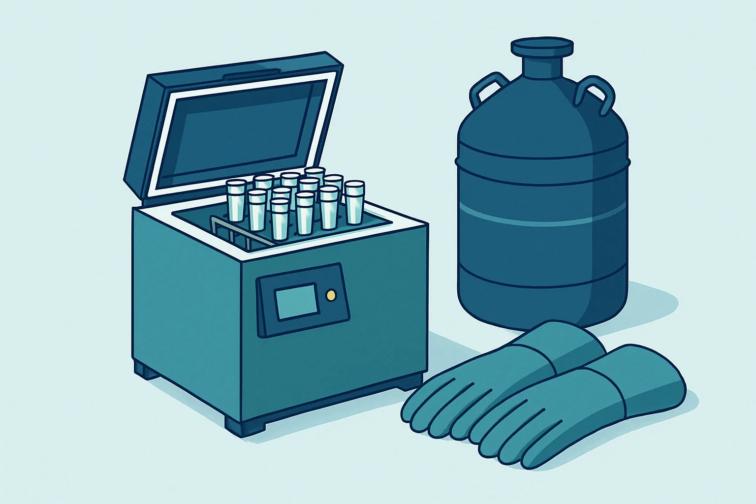
A vial of cells that thaws with poor viability almost never died of cold. Cells tolerate low temperature remarkably well; at liquid nitrogen temperatures essentially all chemistry stops and nothing degrades on any timescale a laboratory cares about. The damage happens on the way down and on the way back up, in a temperature window only a few tens of degrees wide.
Inside that window, water changes phase. Ice forms, and its formation does two quite separate kinds of harm: mechanical disruption from crystals themselves, and chemical injury from the solution that becomes progressively more concentrated as pure water is removed from it. Which of the two dominates depends almost entirely on how fast the temperature falls, and the two run in opposite directions. That opposition is why cryopreservation has an optimal cooling rate rather than a fastest-is-best rule.
Everything else in a protocol, from cryoprotectant choice to container shape, widens the narrow band of conditions in which neither damage type is severe. Knowing what each step protects against turns a recipe into something diagnosable when it fails.
Key takeaways
- Cells are not damaged by cold but by ice formation and by the concentrated solute left behind as water freezes out.
- Fast cooling risks intracellular ice; slow cooling risks osmotic and solute injury. The optimum is a compromise between the two.
- Cryoprotectants work mainly by reducing the fraction of water that turns to ice at a given temperature, not by any antifreeze magic.
- Long-term storage must sit below the glass transition of the solution, which is why vapour phase nitrogen storage is the standard.
- Thawing should be fast, and the cryoprotectant should be removed gently, because rapid dilution causes its own osmotic damage.
What Freezing Physically Does to a Cell
A cell is mostly water containing dissolved salts, proteins, sugars and metabolites, enclosed by a lipid membrane that lets water pass fairly freely and most solutes pass slowly or not at all. When the temperature falls below freezing, the first thing that happens is nothing at all: the solution supercools, remaining liquid several degrees below its equilibrium freezing point because ice needs a nucleation event to begin.
When nucleation finally occurs, it almost always happens outside the cells first. The extracellular volume is larger, contains more particles capable of seeding a crystal, and is not protected by a membrane. Ice grows in the space between cells while the cell interiors remain liquid.
This creates the central problem. Ice is pure water; the salts and other solutes are excluded from the crystal lattice and remain dissolved in a shrinking volume of unfrozen liquid between the growing crystals. That residual solution therefore becomes steadily more concentrated as freezing proceeds. Its osmotic pressure rises far above anything a cell ever experiences physiologically.
The cell responds as any osmotic system does: water crosses the membrane down the gradient and the cell shrinks. This dehydration is not incidental. It is how a cell avoids freezing internally, since a shrunken cell with a concentrated interior has a depressed freezing point and less water available to crystallise. But dehydration carries its own costs. Membrane lipids are forced together, proteins meet salt concentrations that denature them, and the cell can shrink past recovery. So it is caught between two injuries: time to dehydrate brings solute damage, and denying that time leaves water to freeze inside.
Ice Crystal Formation and Membrane Damage
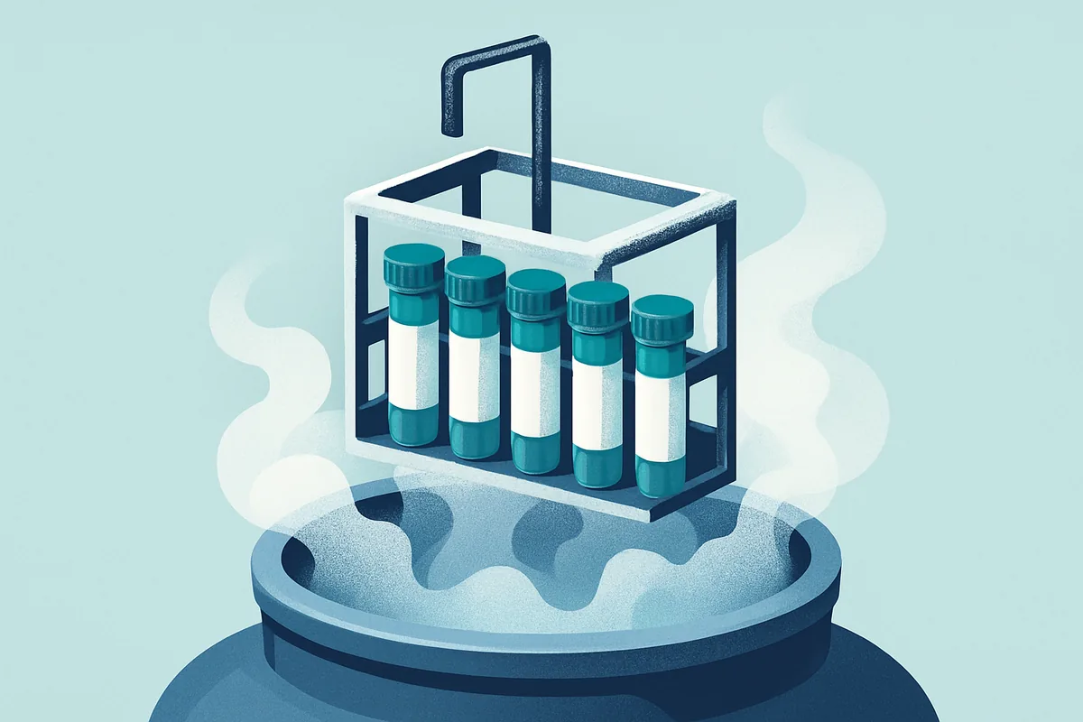
Intracellular ice is generally lethal, though not because crystals stab the cell like knives, an image that persists in teaching but describes the physics poorly. Crystals forming inside a cell nucleate at many points and grow into the cytoplasm, deforming organelle membranes and the cell surface as they expand; mitochondrial and nuclear membranes are as vulnerable as the plasma membrane. Even crystals small enough to be tolerated are thermodynamically unstable, and on warming they recrystallise, with small crystals dissolving and redepositing onto larger ones that have lower surface energy. A cell that survived cooling with a scattering of tiny crystals can be destroyed during a slow thaw as they consolidate.
This is why cooling rate matters so directly. Water leaves a cell at a finite speed set by membrane permeability, which itself falls sharply as temperature drops. If the temperature falls faster than water can exit, the cell interior stays too dilute relative to its surroundings, becomes deeply supercooled, and nucleates internally.
Cell types differ in membrane permeability and surface-to-volume ratio, which is why no single optimal rate exists. Small, highly permeable cells such as red cells dehydrate quickly and tolerate much faster cooling. Large cells with low permeability, oocytes among them, need far slower cooling or an entirely different approach.
| Damage mechanism | Dominant when cooling is | Underlying cause | Typical mitigation |
|---|---|---|---|
| Intracellular ice | Too fast | Water cannot leave the cell in time | Slow the rate, add permeating cryoprotectant |
| Solute concentration injury | Too slow | Prolonged exposure to concentrated salts | Speed the rate, reduce time in the damaging window |
| Excessive shrinkage | Too slow | Osmotic water loss beyond recovery | Balance rate, use non-permeating agents |
| Recrystallisation | Any, during warming | Small crystals consolidating into large ones | Thaw rapidly, avoid partial thaw and refreeze |
| Devitrification | On warming from vitrified state | Glass converting to crystal | Warm rapidly through the transition |
The table makes the shape of the problem clear. Two of the mechanisms are made worse by slow cooling and one by fast cooling, and the remaining two are consequences of how warming is handled. The optimum sits where the two cooling-related curves cross, which for most nucleated mammalian cells in culture is a cooling rate of roughly one degree per minute.
How Cryoprotectants Change the Equation
Cryoprotectants are usually described as substances that stop ice forming. That is misleading. They do not prevent ice; they change how much ice forms at a given temperature and how concentrated the residual liquid becomes.
Permeating agents, of which dimethyl sulfoxide and glycerol are the most widely used, cross the cell membrane and distribute through the cytoplasm. Their main effect is colligative: adding a large amount of any solute depresses the freezing point and, more importantly, means that at any given subzero temperature a smaller fraction of the water has turned to ice. Less ice means a larger residual unfrozen volume, which means the salts left behind are diluted across more liquid and reach damaging concentrations only at much lower temperatures. The cell spends less of its journey in the dangerous window, and it dehydrates less severely.
Both agents also hydrogen-bond extensively with water and interact with membrane lipids, which appears to stabilise membranes during the extreme dehydration of freezing. Dimethyl sulfoxide permeates faster and suits most cultured cells; glycerol crosses membranes more slowly and is favoured for some primary cells, sperm and certain microorganisms.
Non-permeating agents work differently. Sugars such as sucrose and trehalose, and polymers including hydroxyethyl starch, stay outside the cell, raising external osmolality so that water leaves before freezing begins and less intracellular water is available to crystallise. They also appear to protect membranes directly, replacing hydrogen bonds that water normally makes with lipid head groups. Serum proteins in a typical freezing medium contribute similarly.
The costs are real. Dimethyl sulfoxide is toxic at room temperature over anything more than brief exposure, and toxicity rises steeply with temperature and concentration. It also has biological activity of its own, including effects on differentiation in some cell types, which matters when the recovered cells are the experiment. Hence the insistence on adding it cold, freezing promptly, and removing it quickly after thaw.
The One Degree Per Minute Convention
The figure of one degree per minute has the status of folklore, but it is an empirical optimum for a fairly broad class of cells rather than an arbitrary number. It is slow enough that most nucleated mammalian cells can dehydrate as the surrounding solution freezes, and fast enough that they are not held for long in the concentrated solution that dehydration produces.
Three approaches deliver it. A passive freezing container, an insulated block with vial wells placed in a minus eighty freezer, produces roughly the target rate by simple thermal resistance. These are cheap and reliable provided the container starts at room temperature and is not overfilled, since both change the rate. Designs relying on an alcohol bath need the fluid replaced periodically, and a container with degraded fluid cools at the wrong rate while looking identical from outside.
A controlled rate freezer does the job properly, injecting nitrogen vapour under feedback control to follow a programmed profile, and it can compensate for the latent heat of fusion. When a sample nucleates, the phase change releases heat and the temperature briefly jumps back up; a programmable freezer can overshoot downwards just before this point, seeding nucleation at a controlled moment. Uncontrolled, that supercooling and release vary from vial to vial, which is one reason passive freezing gives more variable results across a batch.
The third approach is not to aim for one degree per minute at all. Vitrification uses very high cryoprotectant concentrations and extremely fast cooling, so that the solution passes into a glassy solid without crystallising at all. It is standard for oocytes and embryos, where conventional slow freezing performs poorly, and it demands small volumes and careful handling because the concentrated cryoprotectant is toxic and the cooling must be genuinely rapid.
The rate only needs controlling through the range where phase change and solute effects occur, from a few degrees below zero down to around minus fifty or sixty. Below that, the sample can go into storage without further attention to rate.
Storage Temperatures and the Glass Transition
Once a sample is cold enough, molecular motion becomes so slow that the residual unfrozen solution stops behaving as a liquid and becomes a glass: an amorphous solid with no crystal structure but no meaningful mobility either. The temperature at which this happens is the glass transition, and for typical cryoprotectant solutions it lies somewhere near minus one hundred and thirty degrees.
This number is the single most important threshold in storage. Above it, the residual liquid still moves, however sluggishly: crystals grow, recrystallisation continues, and degradation is retarded rather than stopped. Below it, nothing moves. Liquid nitrogen boils at about minus one hundred and ninety-six degrees, comfortably below the transition, and the vapour above it in a well-designed dewar stays colder than the transition throughout the usable volume. Temperature rises with height in that column, which is why liquid level is monitored so carefully: a tank running low leaves upper racks in the warm part of the vapour, where samples are not obviously ruined but are no longer genuinely stable.
Vapour phase storage is standard partly for this reason and partly because submerged vials can admit liquid nitrogen through imperfect seals. On warming, trapped liquid expands violently and vials can rupture. Immersion also creates a plausible route for cross-contamination where vials from different sources share a tank.
Mechanical freezers running at minus eighty degrees are a different proposition. That temperature is well above the glass transition, so storage there is a holding measure rather than a bank. Viability drifts down over months, and the drift is faster for some cell types than others. A minus eighty freezer is where a vial waits for a few weeks before going into nitrogen, not where a cell line lives.
Thawing Fast and Removing DMSO
Warming is the mirror of cooling, and the rules are not symmetrical. Where cooling should be slow, warming should be fast.
The reason is recrystallisation. Whatever small ice crystals exist in the sample are unstable, and as the temperature rises through the range where molecular mobility returns they consolidate into larger, more damaging ones. Passing through that range quickly gives them no opportunity. A vial moved from nitrogen straight into a water bath at body temperature, agitated gently, and removed the moment the last ice disappears will have crossed the dangerous range in a couple of minutes.
Two errors are common. Leaving a vial in the bath after thawing exposes cells to concentrated dimethyl sulfoxide at the temperature where it is most toxic. And partial thawing followed by refreezing, which happens whenever a rack is lifted from a tank and left on a bench while vials are located, causes exactly the recrystallisation a fast thaw avoids.
Removing the cryoprotectant introduces the last osmotic hazard. The thawed cell contains a high concentration of permeating agent. Dropping it into a large volume of plain medium creates a steep gradient in the opposite direction from freezing, and water rushes in faster than the cryoprotectant can leave. Cells swell, and some burst.
The remedy is dilution in steps, adding medium drop by drop at first so the external concentration falls slowly enough for the agent to diffuse out alongside the water coming in. A non-permeating buffer such as sucrose in the first step holds external osmolality up while the permeating agent departs. Centrifugation removes residual agent where the cell type tolerates it, but spinning cells that have just been frozen is itself stressful, and for delicate primary cells a stepwise dilution with a medium change hours later is gentler.
Viability Testing After Recovery
The number that matters is not how many cells are alive an hour after thaw but how many go on to function. These are different quantities, and the gap between them is where most disappointment lives.
Membrane integrity dyes, of which trypan blue is the oldest and various fluorescent equivalents the more sensitive, report only whether the plasma membrane currently excludes a dye. A cell with irreversible mitochondrial damage, fragmented DNA, or a triggered apoptotic programme will exclude dye perfectly well for hours. Counts taken immediately after thaw therefore run optimistically high, and the deficit shows up a day later as a plate that has not attached or a culture that fails to divide.
Two habits improve the picture. The first is to count later, since a viability assessment taken some hours after plating correlates far better with what the culture will do. The second is to measure recovery rather than viability: the proportion of the cells originally frozen that are alive and attached at a defined point after thaw. That single figure captures losses during freezing, thawing and the first hours of culture, and it is the one worth tracking across batches. Metabolic assays and functional or differentiation tests add more, at the cost of time, and are worth reserving for comparisons between protocols rather than routine checks.
Beyond viability, a cell bank needs identity and cleanliness checks. Cross-contamination between cell lines is a long-standing and well-documented problem in research, and the moment a bank is created is the natural point to confirm that the line is what its label claims and is free of mycoplasma. A carefully frozen bank of the wrong cells is a worse outcome than a poorly frozen bank of the right ones.
Frequently asked questions
Why is my viability high immediately after thawing but low the next day?
Because dye exclusion assays report membrane integrity at the moment of testing, not the cell’s prospects. Freezing injures mitochondria, damages DNA and can trigger apoptotic programmes that take hours to run to completion, and throughout that period the plasma membrane stays intact and the cell counts as viable. The practical fix is to shift your assessment later, counting attached or viable cells some hours after plating, and to treat that delayed figure as the real one when comparing freezing protocols.
Can I freeze cells directly in a minus eighty freezer without a controlled rate device?
You can, but the cooling rate will be uncontrolled and usually too fast, because a vial dropped straight onto a freezer shelf cools far quicker than one degree per minute at the start. An insulated passive freezing container is the minimum sensible measure and gets most laboratories close to the target rate at trivial cost. Whatever the method, minus eighty is a staging point rather than a destination, since it sits above the glass transition and viability declines over months.
How long can cells be stored in liquid nitrogen?
Below the glass transition there is no known mechanism of biological degradation operating on laboratory timescales, because molecular motion has effectively ceased. Practical limits come from everything except the cold: a tank that ran dry unnoticed, vials whose labels became illegible, seals that admitted nitrogen, or inventory records that no longer match reality. Reliable long-term banking depends far more on temperature monitoring, redundant storage in a second location, and disciplined record keeping than on anything about the freezing itself.
Is dimethyl sulfoxide always the right cryoprotectant?
It suits most cultured mammalian cell lines and it is the default for good reasons: it permeates quickly, works at moderate concentrations, and has a long track record. It is not universal. Glycerol suits some primary cells and microorganisms better, sugars and polymers substitute for it where a permeating agent is undesirable, and applications where the cells will be given to a patient or where the agent’s own biological activity would confound the experiment often need alternatives. If your recovered cells behave oddly in ways that track with freezing, the cryoprotectant deserves suspicion.
Does the vapour phase really protect against cross-contamination?
It reduces one specific route rather than eliminating risk. Liquid nitrogen can carry material between containers, and vials with imperfect seals can take liquid in and let it out again, so immersion creates a plausible transfer path that vapour storage removes. Vapour phase does not address contamination introduced during handling, shared racks, or samples that were already contaminated when banked. Segregating untested or known-contaminated material into separate tanks remains the more consequential control.
The habit that most improves outcomes is treating freezing as a designed process rather than an errand at the end of a long day. Cells should be healthy, growing well and harvested in their exponential phase before they go anywhere near a cryovial, because a stressed culture freezes badly regardless of technique. Cryoprotectant should be added cold and the vials frozen promptly rather than left standing. Records should say which passage, which medium, which date, and how many cells went into each vial. And a single vial from every batch should be thawed as a test soon after banking, because a bank whose recovery has never been checked is not a bank but an assumption.
This is education, not medical advice. Laboratory results only carry meaning alongside your symptoms, history and examination. Talk to a qualified clinician about your own results before changing anything about your care or supplements.



