

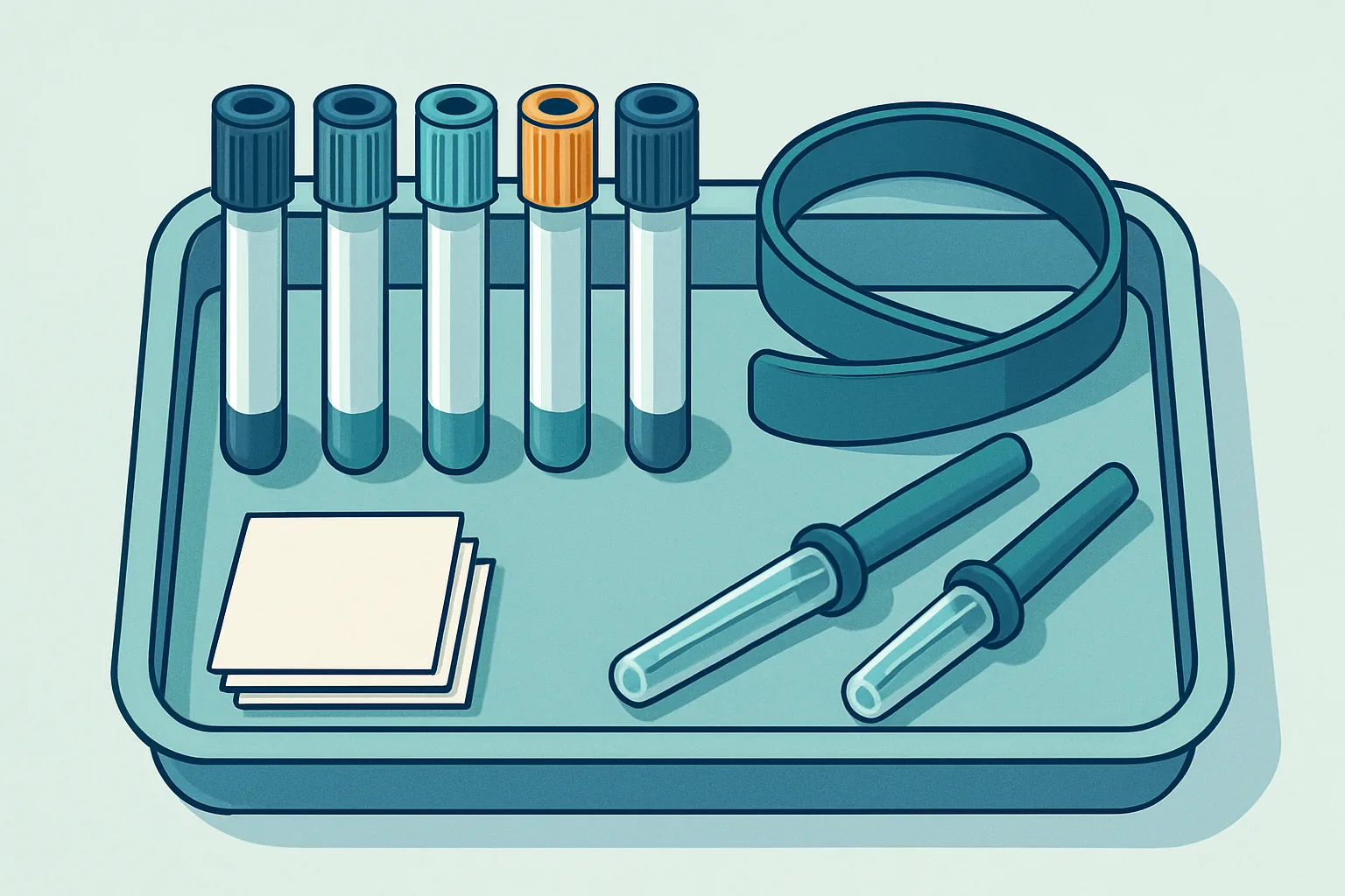
A blood test feels like a measurement of you. In practice it is a measurement of a sample, and the sample is not quite the same thing as the person it came from. Blood behaves differently once a tourniquet is applied, once it has left the vein, and once it sits in a tube with an additive designed for a different purpose.
Most of the time this does not matter, because the changes are small relative to the range of values considered normal. But for a handful of common analytes the changes are large enough to move a result across a decision threshold, and the laboratory receiving the sample usually cannot tell that it happened. The result looks entirely plausible. It is simply wrong about the patient.
Studies of laboratory error consistently find that most mistakes occur before analysis begins, and collection is the largest single contributor. What follows walks through the mechanisms in roughly the order they occur during a draw.
Key takeaways
- A prolonged tourniquet concentrates the blood locally and pushes potassium out of muscle and red cells, raising several results.
- Clenching the fist repeatedly during a draw can raise potassium substantially in a person whose potassium is normal.
- The order in which tubes are filled matters because additives carry over from one tube to the next.
- Too narrow a needle, or a difficult draw, shears red cells and produces haemolysis that corrupts specific analytes.
- Underfilled tubes change the ratio of blood to additive, which invalidates coagulation results in particular.
Why Tourniquet Time Shifts Potassium
A tourniquet works by obstructing venous return while leaving arterial inflow largely intact. Blood continues to arrive in the limb and cannot easily leave, so the veins distend and become easier to puncture. That is the intended effect. Several unintended ones begin almost immediately.
The first is haemoconcentration. The raised pressure inside the distended vessels drives water and small molecules out through the capillary wall into the tissue. Anything too large to follow stays behind and becomes more concentrated. Within a couple of minutes this raises the apparent concentration of proteins, of everything bound to proteins, and of cells, while small freely diffusible molecules such as sodium and glucose are barely affected because they move with the water.
Calcium deserves specific mention because roughly half of it circulates bound to albumin. When albumin concentrates, total calcium rises with it, and a borderline calcium taken after a long tourniquet application can look abnormal when the active free fraction has not changed at all.
The second effect is local metabolic. With venous outflow blocked, the tissue downstream keeps consuming oxygen and works anaerobically. Local pH falls as lactate and carbon dioxide accumulate, and acidosis shifts potassium out of cells into plasma. Lactate itself rises steeply, which makes a tourniquet essentially incompatible with a meaningful lactate measurement.
Both effects grow with time, which is why guidance converges on releasing the tourniquet as soon as blood flow is established and keeping total application to about a minute. The practical difficulty is that the patients with the hardest veins are the ones on whom a tourniquet stays longest, so the error concentrates in exactly the people who are drawn most often.
Fist Clenching and Falsely High Readings
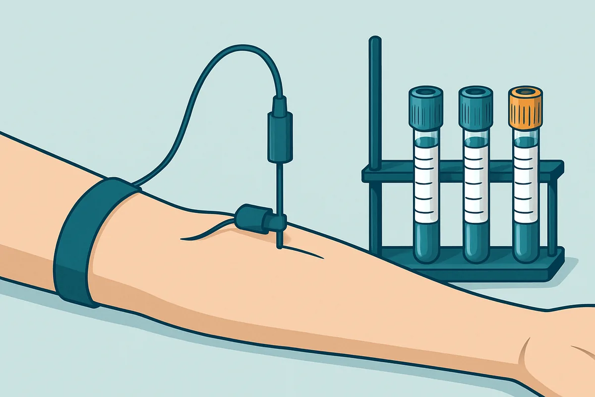
Asking a patient to make a fist to bring up a vein is common and, done once and held gently, largely harmless. Asking them to pump the fist repeatedly is a different matter.
Contracting muscle releases potassium. Skeletal muscle holds a very high intracellular potassium concentration, and each contraction allows some to escape into the interstitial fluid and then into the local venous blood. Because the tourniquet is preventing that blood from leaving the arm, the released potassium accumulates in precisely the vessel about to be sampled. Vigorous pumping over a minute or two can raise the measured potassium by an amount large enough to prompt a repeat sample, an urgent phone call, or in the worst case a treatment decision, in a person whose systemic potassium is entirely normal.
The muscle activity also releases other intracellular contents. Lactate rises, and creatine kinase, an enzyme abundant in muscle, can rise too, which matters because it is used to assess muscle injury.
The correction is straightforward: the patient may clench once to help the vein fill and should then relax. If a vein cannot be raised without pumping, the better options are warming the arm, allowing it to hang below heart level briefly, or choosing a different site.
A related situation produces the same artefact outside the phlebotomist’s control. A patient who has cycled or walked briskly to the appointment arrives with a genuine and transient shift in several muscle-derived analytes, which is one reason a short rest before sampling is recommended for anything involving potassium, lactate or muscle enzymes.
The Order of Draw and Additive Carryover
Blood collection tubes contain different additives, and those additives are not benign if they end up in the wrong tube. During a multi-tube draw, blood passes through a shared needle and holder, and a small amount of additive from one tube can be carried into the next. The standard order of draw exists to make any carryover harmless rather than to prevent it entirely.
The reasoning becomes clear once you know what each additive does. EDTA, used for full blood counts, chelates calcium. Calcium is essential to the coagulation cascade, so even a trace reaching a coagulation tube prolongs clotting times. EDTA binds other divalent metals too, so carryover into a chemistry tube lowers calcium and magnesium, and because EDTA tubes are supplied as potassium salts, the same trace raises potassium sharply. One additive corrupts three separate things.
| Tube type | Typical additive | What it is for | What its carryover damages |
|---|---|---|---|
| Blood culture | Broth, no anticoagulant | Sterile culture | Drawn first to protect sterility |
| Citrate (light blue) | Sodium citrate | Coagulation testing | Dilutes and lowers calcium in later tubes |
| Serum (red or gold) | Clot activator, gel | Most chemistry | Activator can trigger clotting in later tubes |
| Heparin (green) | Lithium or sodium heparin | Plasma chemistry | Inhibits clotting; alters lithium or sodium results |
| EDTA (lavender) | Potassium EDTA | Full blood count | Raises potassium, lowers calcium, prolongs clotting times |
| Fluoride oxalate (grey) | Sodium fluoride | Glucose and lactate | Fluoride inhibits enzymes used in other assays |
The conventional sequence places blood cultures first to protect sterility, then citrate, then serum tubes, then heparin, then EDTA, then fluoride oxalate. The pattern is that the tubes whose results are most easily ruined sit early, and the additives that do the most damage sit late.
One wrinkle deserves a note. When a butterfly set is used and a citrate tube is drawn first, the air in the tubing is pulled into the tube instead of blood, leaving it underfilled. A discard tube is drawn first to prime the line, and skipping it produces a citrate tube with the wrong blood-to-additive ratio.
Needle Gauge, Flow Rate and Cell Damage
Red cells are flexible but not indestructible. Forcing them through a narrow opening at speed subjects them to shear stress, and beyond a threshold their membranes rupture. This is haemolysis, and it is the most common reason a sample is rejected.
Needle gauge is the obvious variable, and it works in both directions. A needle that is too narrow accelerates the blood and increases shear, which damages cells. But a needle that is too wide for a small or fragile vein encourages the vein to collapse under vacuum, causing turbulent flow and intermittent stoppage that also damages cells. A common gauge for routine adult venepuncture balances these, with smaller gauges reserved for difficult veins and paired with gentler collection.
Vacuum is the other major factor. Evacuated tubes pull blood at a rate determined by the vacuum, and a vein that cannot supply that rate will partially collapse. Using a syringe allows the operator to control the pull, but introduces its own hazard: pushing blood through a needle into a tube under pressure shears cells efficiently. Blood should be transferred using a proper transfer device, never forced.
Other contributors are quietly common. Probing to locate a vein causes tissue damage and cell rupture, and drawing through an existing haematoma pulls damaged cells into the sample. Alcohol that has not dried lyses cells at the puncture site. Vigorous shaking of tubes, rather than gentle inversion, breaks cells mechanically.
The consequences fall unevenly. Potassium, lactate dehydrogenase and certain enzymes are concentrated inside red cells at levels far above plasma, so their release raises measured values dramatically. Haemoglobin released into the plasma also interferes optically with assays that measure absorbance, and it interferes chemically with some others. Meanwhile many analytes, including sodium, urea and most proteins, are affected very little.
Underfilled Tubes and Dilution Errors
Every additive tube is designed around a specific fill volume, because the additive quantity is fixed and the ratio to blood is what matters.
Coagulation testing is the strictest case. Citrate tubes contain a liquid anticoagulant intended to be diluted by a defined volume of blood, conventionally nine parts blood to one part citrate. Citrate binds calcium, and the testing process reverses this by adding a fixed amount of calcium back. If the tube is underfilled, the citrate concentration is too high, more calcium is consumed in the reversal, and clotting times are falsely prolonged. Because that prompts dose changes in patients on anticoagulants, coagulation laboratories reject underfilled tubes rather than reporting them with a comment.
EDTA tubes are more forgiving but not immune. Excess EDTA relative to blood draws water out of red cells osmotically, shrinking them. That lowers the mean cell volume and, because haematocrit is usually calculated from cell volume, lowers the haematocrit too. Severe underfilling can also distort cell morphology enough to make a blood film difficult to interpret.
Heparin tubes are the most tolerant, since heparin acts catalytically rather than stoichiometrically, but grossly underfilled heparin tubes still risk micro-clotting that will block an analyser.
The other filling problem is the partial fill caused by loss of vacuum, which happens when a tube is dropped, stored past its expiry, or punctured and withdrawn. A tube that stops filling early during an otherwise good draw should be replaced rather than accepted.
Line Draws and Contamination With Fluids
Drawing from an existing intravenous line or central catheter is tempting, particularly in hospital patients who are being sampled repeatedly. It avoids another needle, and it is the single most productive source of spurious results in inpatient laboratory medicine.
The problem is that a line contains fluid. If a patient is receiving an infusion, blood drawn from that line or from a vein downstream of it is diluted by whatever is running. The characteristic pattern is a set of results that make no biological sense together: glucose in the tens of millimoles because dextrose is running, sodium and chloride that mirror the infusion rather than the patient, and everything else diluted proportionally. A raised potassium in a patient receiving potassium-containing fluid is the version most likely to trigger action.
Lines also retain drugs. A catheter flushed with heparin yields samples with prolonged clotting times unless a sufficient discard volume is withdrawn first. Sampling for drug concentrations through the same line used to administer the drug produces meaningless results, a recurring error in antibiotic monitoring.
The mitigations are procedural. Stop the infusion for a defined period before drawing where clinically safe. Draw from the opposite arm rather than downstream of a running line. Withdraw and discard an adequate volume before collecting, sized to the catheter’s dead space. Never sample a drug from the lumen through which it was given.
The most important defence is scepticism about implausible combinations. A sodium result impossible alongside the patient’s clinical state, or a set of chemistry results all diluted by the same proportion, should prompt a repeat by clean venepuncture.
What a Good Draw Looks Like Start to Finish
Pulling the threads together, a technically sound draw is a sequence of small decisions rather than a single skill.
It begins before the needle. The patient is identified positively and the request checked, including any fasting or timing requirement, since a drug level drawn at the wrong point in a dosing interval is uninterpretable regardless of how well it was collected. The patient is seated or lying and has rested briefly.
Site selection follows. A vein that can be felt is better than one that can be seen, and the antecubital fossa is preferred because the vessels there are large enough to tolerate a standard needle without collapsing. Arms with a fistula, recent surgery, lymphoedema or an active infusion are avoided.
The tourniquet goes on, the vein is confirmed, the skin is cleaned and allowed to dry fully. The needle enters at a shallow angle in one deliberate movement, without probing. As soon as blood flows, the tourniquet comes off. Tubes are filled in the correct order, each allowed to fill to its mark under its own vacuum, and each inverted gently the specified number of times immediately after removal rather than at the end.
The needle is withdrawn, pressure applied, and the tubes labelled at the patient’s side. Labelling away from the patient is how misidentification happens, and misidentification is the one pre-analytical error that no laboratory check can catch, because the sample is perfectly good and belongs to someone else.
Transport comes last and is frequently the weakest link. Samples should reach the laboratory promptly and without violent agitation. Pneumatic tube systems save time and can shear cells if the carrier is poorly padded or the route aggressive. Extremes of temperature should be avoided in both directions, since cold slows the pumps that hold potassium inside red cells and warmth accelerates metabolism.
None of this is exotic, and that is rather the point. The interventions that most improve result quality are unglamorous: release the tourniquet early, do not let the patient pump, fill tubes in order and to the line, choose a sensible needle, avoid lines, label at the bedside, and get the sample to the laboratory quickly. Analysers have become extraordinarily precise, and that precision is wasted on a sample that was already wrong when it arrived. For a patient facing an unexpected result, the useful question is not only what the number means but whether the draw could have produced it, because a repeat sample drawn carefully resolves a surprising proportion of laboratory mysteries at no cost beyond a second needle.
Frequently asked questions
Why was I asked not to clench my fist during the draw?
Muscle contraction releases potassium from muscle cells into the surrounding blood, and with a tourniquet in place that blood cannot leave the arm, so the potassium accumulates exactly where the sample is being taken. Repeated pumping can raise the measured potassium enough to look abnormal in someone whose actual level is normal. A single gentle clench to help the vein fill is fine; sustained pumping is what causes the problem, along with smaller rises in lactate and muscle enzymes.
Does it really matter which order the tubes were filled in?
Yes, because small amounts of additive carry over between tubes through the shared needle and holder. The most consequential example is potassium EDTA from a full blood count tube reaching a chemistry tube, where it raises potassium and lowers calcium and magnesium simultaneously. Carryover into a coagulation tube prolongs clotting times because EDTA removes the calcium those tests depend on. The standard sequence is arranged so that the most easily damaged tubes are filled before the most damaging additives are opened.
Can the laboratory tell that a sample was collected badly?
Sometimes. Haemolysis, clotting, underfilling and gross dilution are usually detectable, and modern analysers measure indices that quantify the degree of haemolysis so the laboratory can suppress affected results. What cannot be detected is a technically perfect sample collected under distorting conditions: a prolonged tourniquet, a clenched fist, a patient who ran up the stairs. Those samples look completely normal to the laboratory and produce plausible numbers, which is why the collection record matters.
Why does a difficult draw so often need repeating?
Difficult draws take longer, which extends tourniquet time and increases haemoconcentration and local potassium release. They often involve probing, which damages tissue and releases cell contents. Slow or interrupted flow into an evacuated tube causes turbulence that shears red cells, and tubes frequently end up underfilled when the vein collapses. Each of these independently raises the chance of an unusable sample, and they tend to occur together, which is why a repeat is so often needed after a struggle.
Is a sample drawn from my drip line as good as one from a fresh needle?
No, and it is a leading cause of misleading results in hospital. Anything running through the line contaminates the sample, producing chemistry values that reflect the infusion rather than the patient, and lines flushed with heparin ruin coagulation tests. An adequate discard volume before collection reduces the problem but does not always eliminate it. Where a decision depends on the result, particularly for potassium, glucose, sodium or drug concentrations, a fresh venepuncture in the opposite arm is the reliable option.
This is education, not medical advice. Laboratory results only carry meaning alongside your symptoms, history and examination. Talk to a qualified clinician about your own results before changing anything about your care or supplements.



